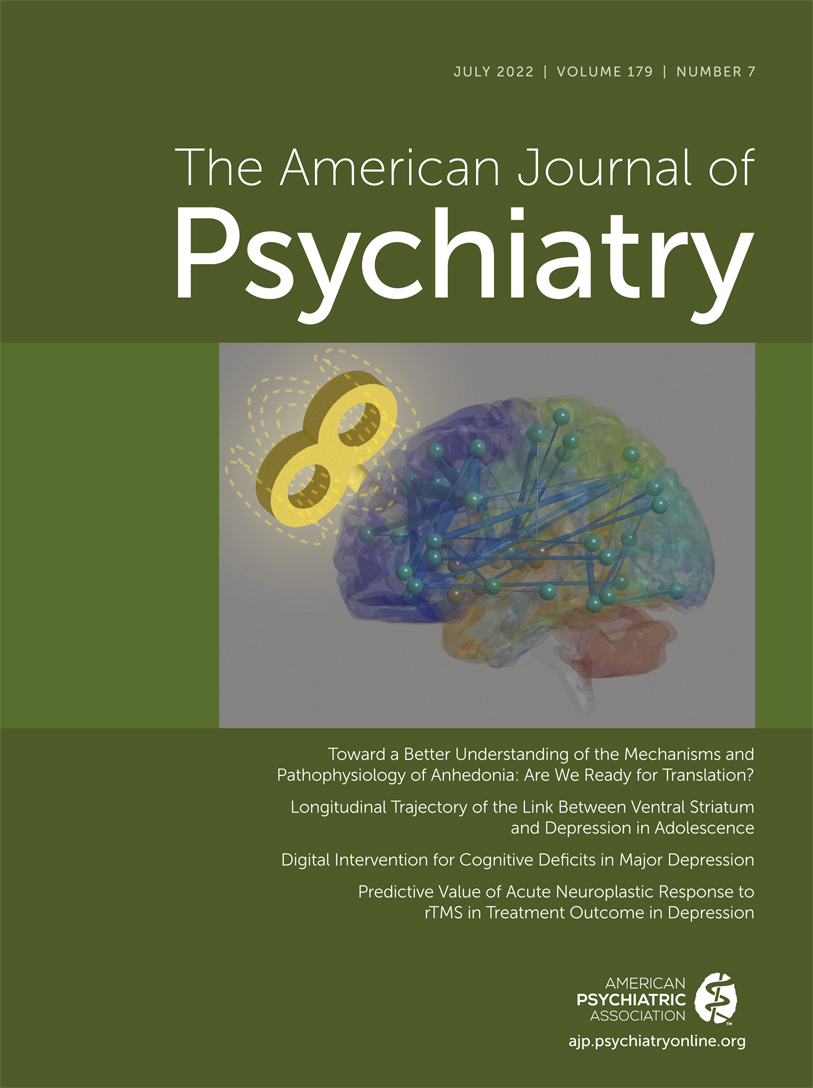
Neuroscientific Advances Supporting New Treatments for Major Depression
This issue of the Journal is focused on major depression. The papers comprising this issue share the ultimate goal of establishing a better understanding of the pathophysiological processes underlying depression with the aim of developing new treatments for depression. Anhedonia, thought to be linked to alterations in reward-related processing, is a hallmark symptom of depression, and we begin the issue with a comprehensive overview on this topic (1). Authored by Dr. Diego Pizzagalli from Harvard Medical School and McLean Hospital, this paper uses two case studies to portray different types of anhedonia, setting the stage for an in-depth discussion of the neural correlates and mechanisms that are associated with reward processes and anhedonia. The overview also summarizes data from treatment studies focused on ameliorating anhedonic symptoms, and importantly emphasizes future directions for developing novel treatments aimed at regaining the ability to experience pleasure, critical for full recovery from the suffering experienced by patients with major depression.
Complementing this review, we include an original research paper that assesses reward-related brain function in adolescents, and the value of characterizing individual differences in reward-related circuit function for predicting longer-term depressive and anhedonic symptoms. Another potentially important finding related to the pathophysiology of depression is presented in a priority data letter demonstrating, in a very large sample, a lack of relation between increased amygdala reactivity and depression. The implications and generalizability of this somewhat surprising finding for hypotheses linking altered amygdala function to depression is thoughtfully discussed in an accompanying editorial. Moving toward the development of new treatments and the ability to predict treatment responses, we present three papers, two of which report results of randomized controlled trials. The first paper assesses the efficacy of a digital intervention for treating cognitive deficits in patients with major depression, and the second compares the efficacy of dextromethorphan plus bupropion to bupropion alone for the treatment of major depression. The rationale for this novel approach is based on using bupropion to increase plasma levels of dextromethorphan, which is hypothesized to work via NMDA antagonism. The final paper in this issue presents data suggesting that characterizing the acute effects of one rTMS treatment on brain function could be useful in selecting depressed individuals that are more likely to respond to a full course of rTMS.
Reward Circuitry Alterations Relevant to Adolescent Depression and Anhedonia
Alterations in reward-related brain responses have been consistently reported in studies of adults with depression, with similar findings reported in a smaller number of studies of depressed adolescents. In addition to depression per se, there has been considerable focus on the relations among hedonic and anhedonic states with dopaminergic function and the function of the reward neural circuitry. The nucleus accumbens, found within the ventral striatal region, is a core component of the reward circuit, which receives input from dopamine neurons that originate from the brain stem ventral tegmental area. Additionally, the ventral striatum receives input, and is interconnected with, other subcortical and cortical regions involved in emotion, reward, motivation, and memory processes. Pan and colleagues (2) use resting-state functional connectivity data from the IMAGEN database to understand the relation between the strength of intrinsic functional connectivity of the ventral striatum to other selected regions of the reward network (i.e., ventromedial prefrontal cortex, presupplementary motor area, anterior insular cortex, posterior cingulate cortex, anterior cingulate cortex, thalamus, and ventral tegmental area [3]) with mood, anhedonia, and depression in adolescents. The IMAGEN consortium is a multisite European study of adolescent brain development related to mental health, and in the current paper the authors report on imaging data and psychiatric symptom assessments that were initially acquired from 303 participants at 14 years of age with follow-up psychiatric assessments 2 and 4 years later (219 participants completed all follow ups). The researchers also assessed brain responses with task-based fMRI to a monetary delayed incentive task. It is noteworthy that in this sample the rate of depressive disorders, which included individuals with subthreshold symptoms, was less than 10% at all time points sampled. Findings revealed that at 14 years of age the degree of connectivity of the right ventral striatal region to other components of the reward network was positively associated with having a depressive disorder. However, this baseline ventral striatal connectivity metric did not predict depression at 16 and 18 years of age. At 14 years of age, anhedonia was found to be correlated with the level of left ventral striatal connectivity with other components of the reward network. Further analyses suggested that baseline ventral striatal connectivity within the reward network was also associated with anhedonia at the follow-up assessments. In relation to the assessment of mood, no significant associations with ventral striatal connectivity were found at baseline and the longitudinal findings were difficult to interpret. These findings derived from resting functional connectivity measures add to existing data linking ventral striatal function to anhedonic symptoms and depression occurring during late adolescence.
The Effects of a Digital Intervention on Depression-Related Cognitive Changes
Cognitive symptoms such as difficulty concentrating, perseverative thinking, slowed cognition, and memory problems are commonly reported by depressed individuals. Additionally, objective data from neuropsychological testing confirms that cognitive alterations exist in subsets of depressed patients. Keefe and colleagues (4) present data from a double-blind randomized controlled trial examining the efficacy of a digital intervention, AKL-T03, aimed at improving cognitive deficits in patient with major depression. This phase 3 trial, building on earlier studies, was sponsored by Akili Interactive Labs, which developed AKL-T03 as a digital tool to help individuals improve their attentional processes. Participants in the study were mildly to moderately depressed, on stable antidepressant medication regimens, and had altered attention and processing speed as assessed with the Brief Assessment of Cognition symbol coding subscale. The experimental and control groups, each consisting of 37 participants, were expected to spend 25 minutes per day, 5 days per week for 6 weeks using either AKL-T03 or a comparator letter/word game. The primary outcome measure for the study was an improvement in sustained attention, and this effect was evident in the AKL-T03 group when compared with the control group. The other measures of cognition that were assessed (e.g., working memory, processing speed, and task switching), as well as the depression measures, did not significantly differ between treatment groups. In their discussion, the authors emphasize the potential clinical utility of this intervention in treating depressed patients with attentional problems, and also address study design issues that may have minimized the likelihood of detecting broader AKL-T03 treatment effects on cognition. In an editorial, Dr. Phil Harvey from the University of Miami discusses the findings from this study, emphasizing the importance of developing treatments focused on depression-related cognitive impairments (5).
Efficacy of a Dextromethorphan-Bupropion Combination in the Treatment of Depression
With the goal of developing more effective and rapidly acting treatments for depression, Tabuteau and colleagues (6) present data from a double-blind randomized controlled trial sponsored by Axsome Therapeutics. The study is interesting as it compares the efficacy of the combination of dextromethorphan and bupropion to that of bupropion alone. Dextromethorphan, well known for its use as cough suppressant, has effects at multiple receptors including its actions as an NMDA receptor antagonist, a sigma 1 receptor agonist, and a serotonin reuptake inhibitor. In this study, the researchers were particularly interested in dextromethorphan’s actions at the NMDA receptor, which follows from data implicating NMDA receptor antagonism as a mechanism underlying ketamine’s rapid antidepressant effects. The logic used in this study for combining bupropion with dextromethorphan is based on bupropion’s CYP2D6 inhibitory effects, an approach aimed to increase plasma levels of dextromethorphan (CYP2D6 metabolizes dextromethorphan to dextrophan). In this relatively small study, participants with major depression were treated over a 6-week period using change in the Montgomery-Asberg Depression Rating Scale (MADRS) as the primary outcome measure to assess efficacy of dextromethorphan plus bupropion to that of bupropion alone. The dextromethorphan/bupropion group (N=43) received 90 mg/day of dextromethorphan with 210 mg/day of bupropion, whereas the bupropion group (N=37) only received bupropion 210 mg/day. Over the course of the study both groups demonstrated decreases in MADRS scores; however, these effects were significantly greater in the dextromethorphan/bupropion group at both 2 weeks and 6 weeks, when compared with the bupropion alone group. Furthermore, remission rates significantly differed between groups such that by end of the study, 46.5% of the participants in the dextromethorphan/bupropion group were deemed to be in remission compared with 16.2% in the bupropion alone group. The most common side effects that occurred in the dextromethorphan/bupropion group were dizziness (20.8% versus 4.2% in the bupropion only group) and nausea (16.7% and 12.5%, respectively). Unlike the psychotomimetic effects that result from ketamine administration, no such effects were reported by the dextromethorphan/bupropion participants. An important issue to keep in mind when interpreting the antidepressant superiority of the dextromethorphan/bupropion combination is that the dose of bupropion used in both groups is less than the 300 mg/day dose that is commonly used to treat depression. In an editorial (7), Dr. Alan Schatzberg from Stanford University considers the interpretation of these findings in relation to the design of the trial and discusses the complexities involved in understanding dextromethorphan’s putative mechanism of action.
Assessing Brain Connectivity Changes to an Initial Session of Repetitive Transcranial Magnetic Stimulation (rTMS) as a Predictor of Treatment Response
Ge and colleagues (8) combine repetitive TMS (rTMS) with fMRI to probe in patients with treatment-resistant depression initial patterns of rTMS-induced brain functional connectivity that may be predictive of subsequent antidepressant responses to a course of rTMS treatment. After an initial 1-Hz rTMS-fMRI assessment, 38 patients were treated with 1-Hz rTMS five times per week over a 4-week period that was applied over the right dorsolateral prefrontal cortical region. Results demonstrated that an initial session of rTMS induced acute reductions in the magnitude of functional connectivity across various brain regions involving different networks. It is noteworthy that these acute changes were transient, as functional connectivity assessed immediately after the initial rTMS session did not significantly differ from pre-rTMS patterns of functional connectivity. By using connectome-based predictive modeling, the researchers were able to predict approximately 30% of the variance in the magnitude of individuals’ treatment responses to the course of rTMS when assessed with the MADRS after 4 weeks of treatment. While the study was performed in a small sample, did not use a sham treatment as a comparator, and was not blinded, these findings provide evidence suggesting that assessing brain connectivity changes to an initial rTMS session may be helpful in identifying individuals that are more or less likely to respond to a full course of rTMS treatment. Dr. Fabio Ferrarelli from the University of Pittsburgh provides an editorial that comments on the implications of these findings in the context of the neuroplastic effects thought to be associated with rTMS treatment (9).
Large-Scale Population-Based Study Does Not Support an Association Between Amygdala Reactivity to Negative Faces and Depressive Symptoms
Numerous fMRI studies have found altered amygdala function to be present in patients with depression. The findings are frequently characterized by increased BOLD signal reactivity in response to the presentation of negative faces or other negatively valenced stimuli as well as in alterations of amygdala resting-state functional connectivity. In a priority data letter, Tamm et al. (10) present data from an extremely large population-based sample from the UK Biobank (N=28,638) that fails to find a strong relation between depressive symptoms and amygdala reactivity in response to the presentation of negative faces. The sample size used in this study is praiseworthy and is consistent with the recent emphasis on the importance of using very large samples for increasing confidence in the reliability of findings that link neuroimaging measures to symptoms and phenotypes (11). However, the applicability of the findings from this study in relation to depressive disorders should be cautiously interpreted. One important issue to consider is that the sample used was not enriched for individuals with depressive disorders, but rather was selected from the overall population. Consistent with this, this sample had relatively low levels of depressive symptoms. In addition, it is important to recognize that the analysis performed in this study examined only one type of response of the amygdala in relation to depressive symptoms, which does not preclude the possibility that other types of stimuli or imaging paradigms might yield more robust associations between depressive symptoms and altered amygdala function. Dr. Alex Shackman and graduate student Shannon Grogans from the University of Maryland, along with Dr. Andrew Fox from the University of California-Davis, contribute an editorial (12) that is very helpful in interpreting these findings in relation to the considerable preclinical and clinical research data implicating involvement of the amygdala in pathophysiological processes underlying depression.
Conclusions
This issue of the Journal presents exciting new data that address treatment issues relevant to major depression as well as to more specific symptoms: depression-related anhedonia and cognitive deficits. Anhedonia and cognitive deficits are of particular interest in treating depression as they are directly linked to the suffering and disability experienced by depressed patients and tend to be less responsive to current antidepressant treatments. The overview on anhedonia by Diego Pizzagalli provides a framework for understanding mechanisms in the brain that mediate reward and reinforcement as well as alterations in these systems that are hypothesized to underly anhedonia. This overview also provides a basis for ideas relevant to new treatments that are specifically focused on reducing anhedonia. Related to this, Pan et al. demonstrate associations between alterations in reward responsive neural circuitry and depression in adolescents, and also suggest that alterations in these systems may help serve to predict anhedonic symptoms over the longer term.
The overview on anhedonia and the paper on neural circuit alterations associated with adolescent depression point to reward-related neural pathophysiological processes relevant to understanding depression. But when thinking about depression, it is also important to consider mechanisms underlying the maintenance of negative emotions and their associated cognitions. In this regard, the priority data letter authored by Tamm et al. presents data from an extremely large, predominantly nondepressed sample that fails to find a strong association between increased amygdala reactivity to negative faces and depressive symptoms. The accompanying editorial by Grogans and colleagues discusses this finding in more depth, and provides a broader perspective supporting the link between amygdala alterations and depression.
The papers in this issue that are directly related to new treatments introduce promising new approaches. Regarding depression-related cognitive dysfunction, Keefe et al. present data supporting the future utility of a digital intervention, which appears to improve sustained attention in mildly to moderately depressed patients that are also being treated with antidepressants. Ge and colleagues provide evidence suggesting the utility of understanding acute brain responses to rTMS at the individual level as a predictor of rTMS antidepressant efficacy. Finally, data from Tabuteau et al. support the efficacy of the combination of dextromethorphan with bupropion in the treatment of major depression.
An imperative for our field is to improve treatment efficacy, remission rates, and the rapidity of responses in patients with major depression. Additionally, the development of reliable predictors of treatment response will allow for matching specific treatments with individual patient characteristics. The papers in this issue are moving us toward these goals by deepening our understanding of the pathophysiological processes involved in depression and importantly, based on neuroscientific advances, laying the groundwork for new promising treatment approaches.
1 : Toward a better understanding of the mechanisms and pathophysiology of anhedonia: are we ready for translation? Am J Psychiatry 2022; 179:458–469 Link, Google Scholar
2 : Longitudinal trajectory of the link between ventral striatum and depression in adolescence. Am J Psychiatry 2022; 179:470–481Link, Google Scholar
3 : The valuation system: a coordinate-based meta-analysis of BOLD fMRI experiments examining neural correlates of subjective value. Neuroimage 2013; 76:412–427Crossref, Medline, Google Scholar
4 : Digital intervention for cognitive deficits in major depression: a randomized controlled trial to assess efficacy and safety in adults. Am J Psychiatry 2022; 179:482–489Link, Google Scholar
5 : Digital therapeutics to enhance cognition in major depression: how can we make the cognitive gains translate into functional improvements? Am J Psychiatry 2022; 179:445–447Link, Google Scholar
6 : Effect of AXS-05 (dextromethorphan-bupropion) in major depressive disorder: a randomized double-blind controlled trial. Am J Psychiatry 2022; 179:490–499Link, Google Scholar
7 : Understanding the efficacy and mechanism of action of a dextromethorphan-bupropion combination: where does it fit in the NMDA versus mu-opioid story? Am J Psychiatry 2022; 179:448–450Link, Google Scholar
8 : Predictive value of acute neuroplastic response to rTMS in treatment outcome in depression: a concurrent TMS-fMRI trial. Am J Psychiatry 2022; 179:500–508Link, Google Scholar
9 : Is neuroplasticity key to treatment response in depression? Maybe so. Am J Psychiatry 2022; 179:451–453 Link, Google Scholar
10 : No association between amygdala responses to negative faces and depressive symptoms: cross-sectional data from 28,638 individuals in the UK Biobank cohort. Am J Psychiatry 2022; 179:509–513 Link, Google Scholar
11 : Reproducible brain-wide association studies require thousands of individuals. Nature 2022; 603:654–660 Crossref, Medline, Google Scholar
12 : The amygdala and depression: a sober reconsideration. Am J Psychiatry 2022; 179:454–457 Link, Google Scholar


