How the Neurocircuitry and Genetics of Fear Inhibition May Inform Our Understanding of PTSD
Abstract
Abstract
Exposure to traumatic events that produce extreme fear and horror is all too common in both military and civilian populations, but not all individuals develop posttraumatic stress disorder (PTSD) as a result of the exposure. What mediates risk and resilience in the development of PTSD and other stress-related psychopathology is of paramount importance to our further understanding of trauma-related psychopathology as well as the development of new treatment approaches. Biological factors, such as genotype and neurobiology, interact with environmental factors, such as childhood background and trauma load, to affect vulnerability and resilience in the aftermath of trauma exposure. One of the core symptoms of PTSD is the inability to control fear, which has led some investigators and clinicians to conceptualize PTSD as a disorder of fear or, more importantly, its inhibition. This review focuses on translational methods that have been used to examine fear conditioning and inhibition of fear in PTSD and summarizes genetic and neurobiological factors related to fear inhibition. The authors also discuss different pharmacological approaches that enhance fear inhibition and may improve treatment outcomes for patients with PTSD.
The popular expression "What does not kill you makes you stronger" points to the fact that some people respond resiliently to trauma. This statement may be true for highly resilient people. However, for those who are vulnerable, a more appropriate statement might be "What does not kill you can make you ill." Such vulnerability is common. Approximately one-tenth of those who survive life-threatening events will develop mental health disorders such as posttraumatic stress disorder (PTSD) or depression or both (1, 2). One of the goals of modern psychiatry is to identify vulnerable individuals and intervene to prevent the development of these disorders by bolstering resiliency. The factors that contribute to resiliency encompass both biological and psychological aspects of the individual as well as the pre- and posttrauma environment (3). It has also been suggested that resiliency is a product of early stress—that is, that resiliency is an adaptive response that maintains homeostasis under stressful circumstances (4). However, this response is true only for some individuals; for others, traumatic stress can increase vulnerability.
Resiliency results from a combination of both biological factors (which are heritable) and environmental factors to which the individual is exposed (Figure 1). The environmental factors that promote resiliency, including social support after trauma, have been the focus of treatment for PTSD. The biological factors may be based on heritable genetic profiles, which may code for neurochemicals and neural mechanisms that promote resiliency. Recent studies have shown that specific gene alleles are associated with resilience, such that even severe levels of child abuse do not result in severe psychopathology (5, 6). The genetic profile may also code for associative learning mechanisms, such as fear conditioning, to enhance fear responses or to enhance fear extinction, which promotes suppression of fear responses to previously fearful stimuli (3).

Vulnerability to the development of PTSD after trauma exposure may be associated with an exaggerated fear response or an inability to control fear responses, which could either be a risk factor for the disorder (7) or an acquired trait of the illness (8). The DSM-IV (9) diagnosis of PTSD requires exposure to a traumatic event and a cluster of symptoms associated with that event (e.g., psychological and physiological reactions to trauma reminders and avoidance of such reminders). Consequently, several theorists (see reference 10, for example) have proposed that conditioning processes are involved in the etiology and maintenance of PTSD. Especially pertinent to this view is the idea that through the processes of Pavlovian conditioning, a neutral (conditioned) stimulus that occurs in temporal contiguity with an aversive (unconditioned) stimulus that innately elicits pain and fear acquires the ability to elicit a fear response in the absence of the unconditioned stimulus. Thus, neutral stimuli (the conditioned stimuli) present at the time of the trauma (the unconditioned stimulus) acquire the ability to elicit a conditioned fear reaction that can be triggered when the person subsequently encounters these or similar stimuli during the course of normal life. Consistent with this hypothesis, emotional and physiological reactivity to stimuli resembling the original traumatic event even years after the event's occurrence is a prominent characteristic of PTSD and has been reliably replicated in the laboratory (see references 11–13, for example). While PTSD is a complex disorder that includes the dysregulation of other emotions besides fear, such as anger or guilt, and is highly comorbid with depression, the study of fear lends itself best to translational approaches (14–16). In this review we first focus on the neural circuits that are involved in inhibition of fear responses and then discuss recent genetic findings in the area. We conclude with a discussion of how these results may be combined in a neurogenetic model that incorporates risk and resilience to trauma-related disorders and indicates prevention and treatment targets in the time course of development of the disorder.
Fear Inhibition as an Intermediate Phenotype
Fear inhibition involves learning of safety signals—that is, the ability to discriminate between danger and safety cues and to suppress fear responses in the presence of safety cues. In the laboratory fear inhibition can be measured by first using a fear conditioning paradigm for fear acquisition, which is then followed by the training of fear inhibition. Fear conditioning is based on a simple Pavlovian conditioning model, in which a neutral conditioned stimulus (CS; for example, a light) is paired with an aversive unconditioned stimulus (US; for example, an electric shock). After a number of pairings, the association is formed so that the CS alone elicits the conditioned response (CR; for example, a fear response). This basic model is used in animal as well as human research to investigate mechanisms of fear acquisition.
Two major laboratory models have been used for behavioral testing of fear inhibition in animals and humans: extinction and conditioned inhibition. While fear acquisition refers to learning that something is dangerous, extinction is a mechanism by which an individual learns that something that previously elicited fear is no longer dangerous—that is, that it is safe. In fear extinction paradigms, a stimulus that was previously paired with an aversive stimulus (the CS+) is then repeatedly presented without the US, so that it no longer elicits a fear response (17, 18). In a basic conditioned inhibition paradigm, the above CS+ pairing is intermingled at the time of training with a separate stimulus (CS–). In other words, the CS– does not co-occur with an aversive stimulus and thus represents safety or inhibition of fear. In another standard conditioned inhibition paradigm, one cue is paired with the aversive stimulus when presented alone (CS+, also referred to as A+) but not when presented in compound with a second cue (CS–, represented as AX–, indicating that the combination of A and X is not reinforced). In this model X should become a safety signal because it signals the absence of the aversive stimulus (19).
In humans, two physiological responses have been used as behavioral outcome measures for fear conditioning: acoustic startle response and skin conductance response. The acoustic startle response is characterized by an integrative reflex contraction of the skeletal musculature in response to a strong stimulus. It provides an excellent model to study emotional processing since the amygdala is directly connected with the startle circuit (16, 20–22). Fear-potentiated startle is the relative increase in the acoustic startle response elicited in the presence of a conditioned stimulus (CS+) that was previously paired with an aversive stimulus (US). The skin conductance response is an index of sympathetic nervous system activity that is frequently used in measuring fear acquisition and extinction in tandem with brain imaging studies using positron emission tomography (PET) or functional MRI (fMRI) (8, 23–26).
Unfortunately, traditional conditioned inhibition paradigms have a number of confounding issues, such as second-order conditioning, external inhibition, and configural learning, that make it difficult to discretely separate excitatory fear learning from inhibition of fear in neural circuits. Myers and Davis (27) developed an animal model using a conditional discrimination procedure that allows for the independent evaluation of excitation and inhibition of fear conditioning. In collaboration with this group, we developed a conditioned inhibition paradigm for use in humans (28) that contains a danger signal (AX+), a safety signal (BX–), and a safety transfer test (combination of A and B, where B reduces fear to A) (Figure 2). The procedure, referred to as a conditional discrimination (abbreviated as AX+/BX–), is based on a paradigm used in earlier learning theory experiments (29, 30). In this experiment, the response to stimulus X is conditional on the presence of either A or B. The A stimulus elicits fear potentiation of startle with training as the subject learns that A and X presented together predicts the US. Stimulus B elicits reduced startle compared to A (i.e., becomes inhibitory) in that B presented with X predicts safety from the US. The presentation of AB results in a reduced startle response compared to the response to A presented with a neutral stimulus because B has become inhibitory.
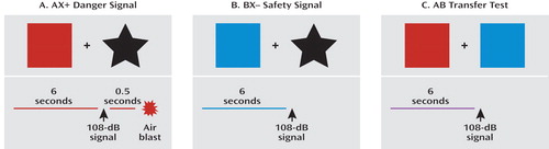
aWhile participating in physiological or neural imaging experiments, the subject observes a computer screen with different colored shapes. In the example in panel A, the danger signal (AX+) is represented by the red square and black star. In panel B, the safety signal (BX–) is represented by the blue square and black star. In panel C, the safety transfer test (combination of A and B, where B reduces fear to A) has both A (red square) and B (blue square) presented simultaneously. The aversive unconditioned stimulus (US) is the air blast that occurs only at the end of the AX+ danger signal. In all three conditions, fear-potentiated startle to the conditioned stimulus (CS) is elicited by a 108-dB startle signal, eliciting an eye blink reflex, which is measured with electromyography.
We translated this paradigm to use in clinical settings and have now demonstrated conditioned inhibition in healthy individuals (28) and in combat veterans with low levels of current PTSD symptoms (31). On the other hand, study subjects with high levels of PTSD symptoms were unable to reduce startle to AB trials (i.e., were unable to transfer fear inhibition). We have also replicated these findings in a sample of veterans with PTSD from the University Hospital Dubrava in Zagreb, Croatia (32), and in a civilian population in Atlanta with high levels of urban trauma (32a). Together these data suggest that PTSD is at least in part a disorder in which inhibition of fear is deficient, even when learned fear in the laboratory is separate from the index trauma(s). An alternative explanation is that fear excitation to the A stimulus in the AB compound is so exaggerated in PTSD that it overwhelms the inhibition from B. This would be consistent with our findings in Vietnam veterans, in that those with the most severe symptoms also had significantly more fear potentiation to the AX+ cue compared to healthy comparison subjects (31). However, our replication samples of combat-related PTSD in Croatia and civilian PTSD in Atlanta did not have increased potentiation to AX+; the group differences were limited to AB and BX– trials, which suggests a selective deficit in fear inhibition.
Both extinction tests and conditioned inhibition focus on active suppression of fear responses through learned safety signals; while fear itself may involve only subcortical areas of the brain located primarily in the limbic circuitry, safety signals may require a cognitive, cortical component (26, 33). This premise is supported by data from our laboratory showing that awareness of the association between the CS and the US is necessary for inhibiting fear responses on the AX+/BX– paradigm (34). Furthermore, a recent study by Weike and colleagues (33) examined the temporal domain of fear conditioning with a danger and safety signal and found that safety signal processing was slower than danger processing. The authors argued that top-down cognitive processes are involved in responses to safety signals, which accounts for the latency in response.
A recent meta-analysis of 15 studies using fear conditioning found that patients with anxiety disorders showed greater levels of fear responses compared to healthy comparison subjects (35). These data suggest that the fear response is overactive or that the inhibition of fear is deficient in PTSD, which has led researchers to use fear conditioning models to examine some of the core PTSD symptoms. One study (36) used a fear-potentiated startle paradigm with veterans diagnosed with PTSD and found equivalent levels of fear potentiation to the danger signal in the PTSD and comparison groups. However, participants with PTSD also potentiated to the safety cue, whereas the comparison subjects did not. Our recent data also show that increased fear responses to safety cues are related to the severity of current PTSD symptoms (31). A recent study of patients with panic disorder (37) found that these patients also had increased fear-potentiated startle responses to the safety cue; this finding may have been related to the patients' increased expectancy of the US during the safety cue. In that study the impaired discrimination between danger and safety appeared to involve both cognitive and physiological deficits. In our study of veterans with PTSD (31), we observed a dissociation between participants' cognitive awareness (they reported that they did not expect to receive an air blast US during the CS– trial) and startle response, which was potentiated in response to the nonreinforced stimulus. On the other hand, a study by Orr and colleagues (38) that used skin conductance to examine fear conditioning in PTSD patients found that patients discriminated between the danger and safety cues better than did comparison subjects. In another study (39), similarly enhanced conditionability in PTSD patients was found when trauma-related cues were used as the US in fear conditioning; the enhanced fear conditioning was also related to slower extinction. Deficient fear extinction in PTSD has been found in several studies that used skin conductance as the physiological measure (26, 39, 40). A recent study of combat-exposed Vietnam veterans and their non-combat-exposed twins (41) found that combat-exposed veterans with PTSD did not have impaired extinction learning but rather had less extinction retention on the day after acquisition and extinction compared to exposed veterans without PTSD. Furthermore, impaired retention of extinction appeared to be an acquired trait related to the disorder since the twins of the veterans with PTSD did not show the same impairment.
While some data with combat veterans suggest that impaired fear inhibition may be an acquired trait (41) associated with current symptom severity (31), other studies have reported that heightened fear responses and decreased inhibition of fear may be predictors of the disorder. A prospective study of police academy cadets (42) found that greater skin conductance responses to threatening stimuli and slower habituation prior to trauma exposure were predictive of PTSD symptom severity after trauma exposure. A similar prospective study with firefighters (7) found that reduced extinction of fear-conditioned responses before the index trauma explained almost one-third of the variance in PTSD symptom severity in later traumatized individuals. It is possible that a decreased ability to inhibit fear is a risk factor for developing PTSD and contributes to the maintenance of the disorder, while decreased extinction retention is a state resulting from the disorder—given that these fear-inhibition phenotypes may have different neural underpinnings, this would explain the above studies.
For the purposes of this review, we will not define fear inhibition as either a vulnerability or an acquired trait of the disorder; more research is needed before such a determination can be made. However, the issue of whether it is a predisposition or a part of the PTSD syndrome itself does not dismiss the utility of impaired fear inhibition as a phenotype. With the development of new techniques for studying fear acquisition and fear inhibition in animal and human subjects, we can begin to understand how the neurobiology of fear is altered in PTSD. Below we review the animal and human data for some of the primary structures involved in fear conditioning and fear inhibition.
Neurocircuitry of Fear Inhibition
The Amygdala
The amygdala, part of the limbic system located in the temporal lobe of the brain, is an integral part of the fear circuitry (43–45). The amygdala comprises several nuclei, which can be roughly divided into the central nucleus and the basolateral nucleus, among several others (Figure 3). Animal studies have shown that the different nuclei function in different ways. For instance, the central nucleus regulates many aspects of the fear response, including the release of cortisol through the paraventricular nucleus of the hypothalamus, increase in startle response via the pons in the midbrain, and modulation of the autonomic nervous system through the lateral hypothalamus (46). Lesions of the central nucleus eliminate fear-conditioned responses, such as fear-potentiated startle (47) and freezing (43), in rodents. The basolateral nucleus projects to the central nucleus and appears to be the locus for associations between the CS and US that result in the acquisition of fear (48).

aPanel A is a schematic diagram illustrating the interaction of the basolateral nucleus (BLA) and central nucleus (CeA) of the amygdala with modulatory regions such as the medial prefrontal cortex (mPFC). The basolateral nucleus is thought to compare conditioned stimulus (CS) inputs and unconditioned stimulus (US) inputs regulating central nucleus activation of the hardwired fear and stress circuitry, leading to inhibition or activation of the fear response. Panel B illustrates recent research that has begun to determine the role of inhibitory neural circuitry in modulating the fear response at the cellular level (49, 50, 53, 54). Sensory inputs as well as associative inputs from the hippocampus and cortex project directly and indirectly to the central nucleus. "On" and "off" inhibitory circuits within the central nucleus are thought to differentially modulate fear output and extinction of fear. Additionally, direct projections from the infralimbic region of the medial prefrontal cortex activate inhibitory neurons in the intercalated region between the basolateral and central nuclei, serving to inhibit, in a top-down manner, the fear output of the central nucleus.
More recent work has begun to outline the cellular microcircuitry that underlies fear expression and inhibition (49, 50). As illustrated in Figure 3B, sensory and associative information projects directly and indirectly to the lateral and basolateral amygdala. Excitatory information from the basolateral nucleus is thought to be gated via inhibitory inputs at the level of the intercalated nuclei situated between the basolateral and central nuclei (51). These inputs are regulated via the medial prefrontal cortex and are thought to be required for extinction of fear (52, 53). Within the central nucleus, information flow is gated by "off" and "on" inhibitory neural networks that are thought to differentially regulate fear expression or inhibition (50, 54). Together, these data suggest that complex inhibitory neural circuitry controls fear behavior and its inhibition, which is dysregulated in pathological states that are marked by amygdala dysfunction.
The bed nucleus of the stria terminalis is a related part of the "extended amygdala" that appears to be associated with nonspecific fear, such as anxiety, which is unrelated to a predictable danger cue used in fear conditioning. Lesions of this structure eliminate anxiogenic effects of bright lights and corticotropin-releasing factor infusions in rodents (55). This region is hypothesized to be more involved in general, nonspecific anxiety and depression symptoms, whereas the central nucleus is thought to be more involved in fear, panic, and cue-specific stress responses.
In accord with animal research, brain imaging studies with humans have found that the amygdala modulates the fear response: left hemisphere damage in temporal lobectomy patients results in loss of fear-conditioned startle (56). In healthy intact humans, several studies using PET and fMRI have shown that presentation of fearful stimuli results in amygdala activation. The stimuli include fearful faces (57, 58) and conditioned fear cues (23, 25, 26). In one study (59), participants were instructed to expect a shock when the "threat" cue was on; that study also found amygdala activation in the threat compared to the safe condition. In a review of 55 imaging studies of the functional neuroanatomy of emotion (60), 25 studies related amygdala activation to fearful stimuli while four studies found activation to positive stimuli. These findings indicate that the amygdala plays an extensive role in regulating the fear response in humans as well as animals.
A preponderance of neuroimaging data from the past decade demonstrate that PTSD patients appear to have greater amygdala activation relative to comparison subjects (see reference 61 for a recent review). PET studies using combat scripts (62) and images (63, 64) and single photon emission computed tomography studies comparing responses to combat sounds and to white noise (65) found greater levels of amygdala activation in PTSD patients. Furthermore, recent fMRI studies have found that trauma-relevant words increase amygdala activation (66). This increased fear response extends beyond trauma-specific imagery: fearful faces also activate the amygdala in PTSD patients more than in comparison subjects (67, 68).
The Prefrontal Cortex
The prefrontal cortex has long been thought to play a role in behavioral inhibition. Nearly two decades ago, animal studies showed that lesions of the medial prefrontal cortex prior to original fear conditioning retard extinction to a tone (69). More recent studies have demonstrated that neurons in the prefrontal cortex may have inhibitory action on the amygdala (24, 70). Just as the amygdala has many subparts, so the prefrontal cortex can be subdivided into the medial and orbitofrontal prefrontal cortex. The anterior cingulate cortex, which is also part of the prefrontal cortex, has both ventromedial and dorsolateral components, which may play different roles in the expression and inhibition of fear, as will be discussed in greater detail below.
A study by Milad and Quirk (71) suggests that the rodent neuroanatomical analogue to the medial prefrontal cortex, the infralimbic prefrontal cortex, has enhanced activity following learning of an extinguished CS. Enhanced infralimbic prefrontal cortex activity was also shown to inhibit the fear response. These authors hypothesize that during the consolidation of extinction, a circuit running from the basolateral nucleus to the prefrontal cortex and back to inhibitory neurons within the amygdala may be strengthened such that when the extinguished CS is reexperienced, the infralimbic prefrontal cortex will represent a feed-forward inhibitory projection that will compete with the fear pathway represented within the basolateral-to-central nucleus projection (Figure 3) (72, 73). The preponderance of evidence indicates that neural plasticity within the amygdala and possibly within the medial prefrontal cortex occurs during the consolidation of extinction learning. As we have seen with studies of consolidation of fear conditioning (74–76), other molecular systems and brain regions, including sensory areas and associative cortical and subcortical areas, are undoubtedly also involved.
Neuroimaging studies in humans have used several paradigms that activate the prefrontal cortex, ranging from simple inhibition of a motor response, such as pressing a button, to more complex tasks in which the subject is required to suppress a response on a cognitive interference task. A simple task may involve responding to a letter when presented alone and withholding a response when the letter is paired with another letter or is shown with a colored background. This type of task is often referred to as a go/no-go task (77). A well-known and frequently used example of a complex task is the Stroop effect, where the meaning of a word (such as the word "red") is in conflict with the color in which it is shown (for example, in blue ink). A novel example of this type of task is the multisource interference task, developed by Bush and Shin (78); in this task the number presented is in conflict with the position in which it is presented so that the subject is required to ignore the interfering information in order to correctly complete the task. When used in an fMRI procedure, this task reliably activates the dorsal anterior cingulate cortex (79).
While the above-mentioned tasks require cognitive inhibition, they do not necessarily involve suppression of emotion and thus may not necessarily map onto the fear inhibition circuitry. A more appropriate task to assess fear inhibition is the emotional Stroop test, in which the emotional content of a word competes with the cognitive content and must be ignored. This task also activates the anterior cingulate, but in a different area than the strictly cognitive interference tasks (80, 81). Emotionally relevant stimuli appear to be processed by the rostral or subgenual area of the anterior cingulate (68), which is anterior to the genu of the corpus callosum, while the dorsal region of the anterior cingulate appears to be more relevant for cognitive tasks (79).
Neuroimaging studies using fear conditioning paradigms demonstrate that fear acquisition and fear extinction also activate the prefrontal cortex, specifically the ventromedial prefrontal cortex (24). Recent developments in the spatial resolution of neuroimaging techniques have resulted in more fine-tuned examinations of this area of the brain. As mentioned above, the rostral or subgenual regions of the anterior cingulate are activated during the presentation of emotional stimuli; these areas are also activated during the regulation of fear (24, 82). Several lines of evidence suggest that this region of the ventromedial prefrontal cortex is associated with inhibition of fear: fMRI data indicate increased activation during extinction recall after extinction learning (24, 83). The ventromedial prefrontal cortex is also activated during fear reversal tasks in which the CS contingencies are switched after acquisition so that a previously conditioned danger cue (CS+) becomes the new safety cue (CS–) (82). Morphometric data show that the thickness of this cortical tissue is correlated with extinction retention (84). Furthermore, the blood-oxygen-level-dependent signal measured with fMRI is greater in the prefrontal cortex when subjects are instructed to "reappraise" a fearful cue—that is, when they actively suppress negative thoughts (85). While functional and morphometric data point to the rostral anterior cingulate during fear inhibition, such data on the dorsal region of the anterior cingulate suggest that this area is associated with fear acquisition (8). Given that this area is also implicated in cognitive tasks (79), it may be activated during the learning procedure of acquisition rather than the fear itself.
Exaggerated fear responses observed in PTSD and the impaired inhibition on conditioned inhibition tasks may be due to a weakened inhibitory control of the amygdala by the prefrontal cortex. PET studies of patients with PTSD show lower activation of the anterior cingulate cortex in response to the emotional Stroop task (86); however, PTSD patients have normal prefrontal cortex activation to nonemotional interference tasks (79, 86). On the other hand, a recent fMRI study of PTSD patients during acquisition, extinction learning, and extinction recall 24 hours later found increased amygdala activation in PTSD patients relative to comparison subjects during extinction learning, and decreased hippocampus and ventromedial prefrontal cortex activation during extinction recall (86a). Weakened prefrontal cortex control of the amygdala may be a risk factor for psychopathology; a recent study of children with depressed parents found a lack of anterior cingulate cortex activation to the emotional Stroop (87).
Interestingly, while all studies found amygdala activation in their study samples as a whole, there were also several instances of individual variability. For instance, LaBar et al. (23) found increased amygdala activation in seven of 10 subjects during early acquisition; Knight et al. (88) found increased amygdala activity only in those individuals who also showed an increased skin conductance response to the danger cue. These individual differences may be due to genotypes that would increase vulnerability to fearful stimuli (89). A recent study found greater fear-potentiated startle in individuals with the short allele of the serotonin transporter gene (90). A gene-by-environment interaction whereby a vulnerable individual is exposed to extreme trauma could lead to the development of PTSD.
Molecular and Genetic Mechanisms of Fear Inhibition
On a molecular level, fear conditioning involves new learning mediated by synaptic plasticity in the amygdala. Both associative fear conditioning and extinction of conditioned fear, a learning process by which a CS is no longer associated with the US, are dependent on activation of glutamate N-methyl-D-aspartate (NMDA) receptors. Administration of NMDA receptor antagonists either systemically (91, 92) or by direct infusion into the basolateral nucleus (93, 94) prior to extinction training blocks the extinction of fear memories. In addition, blockade of NMDA receptors after extinction training also impairs extinction, which suggests that NMDA receptors participate in the consolidation of extinction memories (95). In addition to these data, there is evidence that voltage-gated calcium channels are involved in mediating calcium-dependent synaptic plasticity, which may underlie extinction (96, 97). Additionally, a significant amount of data implicate brain-derived neurotrophic factor (BDNF) in the plasticity underlying fear and extinction learning through its TrkB receptor (74, 98).
There are also substantial data indicating that regulation of the inhibitory neurotransmitter γ-aminobutyric acid (GABA) is altered differentially in the acquisition of fear versus extinction. Injection of an inverse agonist, FG7142, which blocks GABA function, was shown to block the context-specific effects of extinction learning (99). Gephyrin, a scaffolding protein involved in GABA insertion into the surface membrane, is decreased at the protein and mRNA level in the amygdala following fear learning and is increased in the amygdala with extinction learning (100, 101). These data are consistent with enhanced amygdala excitability with fear learning and an increase in amygdala inhibitory tone following extinction. One study (102) demonstrated that blockade of GABA insertion within the amygdala impairs extinction of conditioned fear. Overall these data suggest that modulation of GABA-ergic microcircuitry within the central and basolateral nuclei is critically involved in the regulation of fear and its inhibition with extinction learning.
These molecular data suggest that expression of genes associated with neural plasticity (e.g., BDNF and glutamate receptors), neural inhibition (e.g., GABA receptors and cannabinoid receptors), and stress responsiveness (e.g., glucocorticoid receptors and corticotropin receptor) may be associated with the learning of extinction or impaired fear inhibition. Abnormal fear acquisition or inhibition, as described above, appears to be associated with PTSD, either as vulnerability factors preceding trauma exposure or as a consequence of trauma-related fear conditioning.
To date, much of the research on the genetic basis for PTSD has been gathered via twin studies. Data from these studies indicate that heritability accounts for 30%–40% of the variance in risk for PTSD (103–105). Despite known genetic contributions to risk for PTSD, there have been no linkage studies and only a handful of candidate gene association studies examining genetic main effects to date. In general, these studies have revealed that there are complex interactions between genetic and environmental factors and that many of the identified genetic polymorphisms are in the regulatory promoter regions and not necessarily in the coding regions (106, 107).
Several recent reviews have examined the genetics of PTSD (108, 109, 110), so we will not go into detail on this issue here. No genes have yet been reported that appear to have large main effects across the expected several replications in association with PTSD. Among the more replicated findings to date showing an effect are genes encoding the dopamine receptor 2 (111, 112) and the serotonin transporter (113, 114). However, these findings have not been uniformly replicated (115). As for the genes described above, although two studies have reported no associations with BDNF (116), there have been no reported studies, to our knowledge, of glutamatergic plasticity-related genes, and only one study has sought genetic links between GABA and PTSD (117). In sum, little is known about the genetic mechanisms of PTSD, including the potential genetic role of the glutamatergic and GABA-ergic systems.
Gene-by-Environment Interactions in PTSD
Although a small but growing body of psychiatric research has identified gene-by-environment interactions predicting other mental disorders or associated symptoms, to date only a handful of published studies have presented data on a gene-by-environment interaction predicting PTSD. The first focused on the serotonin promoter length polymorphism (5-HTTLPR), which was originally described as interacting with level of prior stress to predict adult depression (118). Kilpatrick and colleagues (119) identified a gene-by-environment interaction predictive of PTSD in an analysis of individuals exposed to hurricanes in south Florida. The study also classified PTSD patients according to the degree of social support available following trauma exposure. As part of the same hurricane study, Koenen and colleagues (120) reported that the "s" allele of the 5-HTTLPR polymorphism was associated with a lower risk of PTSD in low-risk environments (low crime and unemployment rates) but a higher risk of PTSD in high-risk environments. These results suggest that social environment modifies the effect of 5-HTTLPR genotype on PTSD risk.
The other primary series of studies exploring gene-by-environment effects examined FKBP5, a protein that modulates the glucocorticoid receptor (GR). FKBP5 is a co-chaperone that regulates GR sensitivity (121). Its binding to GR decreases nuclear translocation and decreases GR sensitivity. FKBP5 mRNA and protein expression are induced by GR activation, providing an ultrashort feedback loop for GR sensitivity. Polymorphisms in FKBP5 have been shown to associate with differential up-regulation of FKBP5 following GR activation and with differences in GR sensitivity and stress hormone system regulation. Because of the known role of abnormal GR sensitivity in PTSD (122, 123), FKBP5 appeared to be a good candidate gene that may underlie this component of PTSD and perhaps risk for PTSD. Additionally, FKBP5 polymorphisms had been reported to associate with peritraumatic dissociation (a known risk factor for PTSD) in medically injured children (124). Finally, FKBP5 blood mRNA levels have been found to differentially associate with PTSD in two separate studies (125, 126). In the largest candidate gene study of PTSD to date (5), our group showed a gene-by-environment effect of the polymorphisms in FKBP5 with a history of childhood maltreatment to predict level of adult PTSD symptoms in a traumatized civilian sample. Notably, no main effects were found for the FKBP5 genotype directly associating with PTSD symptoms, nor was there an effect of the gene interacting with adult trauma levels. These data suggest that the interaction of trauma, perhaps during a developmentally critical period, with the hypothalamic-pituitary-adrenal (HPA) axis stress-related genes alters amygdala regulation of fear and its inhibition later in life. These early developmental alterations would then increase risk or enhance resiliency in relation to the development of PTSD in adults following an index trauma.
Temporal Model of Pathophysiology and Treatment Options for PTSD
Conceptualizing PTSD as a disorder of fear conditioning (10, 127–129) leads to the use of fear inhibition experiments to identify vulnerable and resilient individuals and can be used for testing treatments that bolster resilience. Figure 4 shows a putative timeline for the development of fear-related disorders, including PTSD. It starts with factors that increase vulnerability, such as genotype (for example, AA genotype of the FKBP5 rs9296158 gene) and early life stress (such as childhood abuse). Exposure to trauma results in an association of all the external and internal stimuli present at the time of the event and the emotions of fear, horror, and helplessness that define the criteria of trauma. In an individual who will develop PTSD, these stimuli may later serve as reminders and cause symptoms of reexperiencing and hyperarousal. It is possible that intervention at this time, immediately following the event, can prevent the formation of a strong fear association and thus promote recovery. Several studies are under way in which victims of assault are treated in a hospital emergency department within hours after trauma exposure (130).

aThe strength and regulation of fearful memories is affected by numerous factors both before and after the traumatic or fearful event occurs. Genetic heritability comprises up to ∼40% of the risk for both depression and PTSD, and early childhood abuse is a strong risk factor for all mood and anxiety disorders. Further understanding of the roles of genes and environment may allow enhanced prediction of risk and enhancement of resilience in vulnerable populations. Memories are not permanent at the time of the trauma, and psychological and pharmacological approaches to prevent the initial encoding of the trauma are under study. Memories then undergo a period of consolidation in which they shift from a labile state to a more permanent state. Impairing the consolidation (or even reconsolidation) would be an alternative way to prevent the sequelae of long-term trauma memories. The expression of traumatic memories, which can be the source of symptoms in fear-related disorders, is diminished by the process of extinction when repeated therapeutic exposures to the fear-related cues reduce or inhibit the fear memories over time. In contrast, there is some evidence that in individuals who develop PTSD and other pathology, a combination of avoidance of sufficient exposure with intrusive and uncontrollable memories leads to sensitization of the fear response. Enhancing discrimination and extinction of fear memories is a key aspect of recovery in the psychotherapeutic approaches to treating PTSD.
Once the fear memory is formed, it can still be modified by methods that interfere with fear memory consolidation and its potential reconsolidation (131). Consolidation and reconsolidation refer to the phenomenon in which a memory is repeatedly strengthened when trauma reminders become associated with hyperarousal symptoms; this results in a vicious circle by which fear memories lead to anxiety disorders. At this time point, it may be possible to intervene and modify the memory by associating it with safety rather than danger cues (132, 133). If the fear memory is consolidated, trauma reminders will elicit expression of the fear response—that is, amygdala hyperactivity (61). Exaggerated amygdala activity will be evident in the form of symptoms such as intrusive memories, nightmares, exaggerated startle responses (9), and sympathetic nervous system activation, which increases heart rate, respiration rate, and sweating (134). At this point in the course of the development of the disorder, individuals with healthy fear inhibition neurocircuitry might engage the prefrontal cortex to dampen amygdala activity. In fact, a new study (134a) using an extinction paradigm that combined a single reactivation trial 10 minutes prior to extinction found that the fear memory was significantly reduced. More importantly, even a year after extinction, the fear memory was still reduced, indicating resistance to spontaneous recovery of fear. The authors argued that the fear memory, once reactivated, did not have an opportunity to be reconsolidated because of extinction. Treatments that employ mechanisms of fear inhibition, such as extinction, can potentially strengthen the inhibitory controls of the prefrontal cortex on the amygdala, thereby promoting recovery from PTSD.
Although most of this review has focused on mechanisms of fear inhibition and extinction, the failure to extinguish occurs when the excitatory memory of the trauma/CS+ outcompetes the inhibitory memory. Resistance to extinguishing in PTSD may therefore be due either to abnormally strong excitatory conditioning during acquisition or to impaired inhibitory conditioning during extinction. In sum, the confounding contribution of inhibitory and excitatory processes to abnormalities in conditioned inhibition and extinction in PTSD is critical. Figure 4 illustrates how differential memory processes can both inhibit and excite fear memory expression, with PTSD as a potential pathological outcome.
A type of treatment for PTSD that has its basis in extinction is exposure therapy. The term exposure therapy refers to several behavioral and cognitive-behavioral treatment programs that involve confronting feared but safe thoughts, images, objects, situations, or activities in order to reduce pathological (unrealistic) fear, anxiety, and anxiety disorder symptoms. In the treatment of PTSD, exposure therapy usually involves prolonged imaginal exposure to the patient's memory of the trauma and in vivo exposure to various reminders of the trauma. This basic prolonged exposure protocol has been found to be highly effective in the treatment of women with PTSD following physical and sexual assault compared to waiting list or minimal attention control conditions (135–138). Similar exposure therapy programs have been successful with different trauma populations (139–141). Note that there are caveats to data on the prolonged exposure approaches to psychotherapy, however. Studies have often used limiting exclusion criteria and failed to address polysymptomatic presentations, which may render generalizability to a broader population of PTSD patients difficult to determine (142).
Foa and Kozak (143) suggested that two conditions are necessary for emotional processing to occur: activation of the fear memory and the incorporation of corrective information (e.g., that the feared consequence does not occur). These two conditions are met in exposure therapy when the patient confronts actual fear-related stimuli (in vivo exposure), intentionally creates an image of the feared situation, or intentionally retrieves a memory of the traumatic experience (imaginal exposure) and experiences the associated fear reactions (indicated by self-reports of distress or physiological signs of arousal) but in the absence of the feared consequence (e.g., being assaulted). These processes are basically the same as those that occur in extinction, which can be studied directly in animals. Thus, repeated presentations of the CS (in vivo exposure—extinction training) typically elicits the fear response (activation), which then diminishes over the course of repeated trials within an extinction session as well as over the course of successive extinction sessions. Another similarity across exposure therapy and extinction training is a partial return of the conditioned response following exposure/extinction training at the beginning of the next session, referred to as the return of fear (144) in the clinical literature and spontaneous recovery in the extinction literature. In addition, fear often returns in patients who undergo a subsequent major life stress (reinstatement) or even a change in context (renewal).
Pharmacology of Fear Inhibition
As the time course model for PTSD indicates (Figure 4), there may be several points at which therapeutic interventions can be done to prevent the development of PTSD. In addition to psychotherapy, several pharmacological approaches have been used at these time points. For instance, some data suggest that the administration of propranolol in the immediate aftermath of trauma may prevent fear consolidation (145). Studies with animal models have shown that propranolol interferes with the formation of emotional memories (146). Similarly, an early-intervention study of patients in an emergency department suggested that propranolol reduced the development of PTSD symptoms (145). If replicated, these results may provide an exciting approach to preventing PTSD if early intervention is possible, although the time window for response may be limited. However, in most cases, too much time may have passed between the trauma and the treatment, particularly in cases where the trauma occurred in childhood or in combat, when immediate treatment was not available. In such cases, the fear memory is fully consolidated and administration of propranolol would no longer be effective. The most appropriate treatment in these cases would involve exposure therapy, which strengthens fear inhibition through extinction. Pharmacological agents that enhance safety learning would have an important application in this treatment approach.
A recent study showed that D-cycloserine, a partial NMDA agonist, facilitated extinction when rats were tested drug free the next day (147). These results have been fully replicated using freezing as the measure of conditioned fear, even when D-cycloserine is administered up to 4 hours after extinction training (148). This finding led to the first successful clinical test of combining D-cycloserine with exposure-based psychotherapy (149), a result that has since been replicated in several other studies (150–153). Remarkably, the rodent studies have shown that D-cycloserine also seems to block later reinstatement (154). In addition, D-cycloserine leads to generalized extinction (155), where extinction training to one cue in the presence of D-cycloserine leads to a reduction of fear to another CS previously paired with the same US. This could be significant clinically because combining D-cycloserine with exposure-based psychotherapy to specific cues associated with the original trauma might generalize to other cues associated with that traumatic event, even though these are not dealt with explicitly during therapy. A recent meta-analysis of more than 40 animal and human trials examining D-cycloserine and extinction or exposure therapy concluded that D-cycloserine "is a useful target for translational research on augmenting exposure-based treatment via compounds that impact neuroplasticity" (156). Although no studies of the effects of D-cycloserine in exposure therapy for PTSD have yet been completed, several are under way.
The pharmacological enhancement of extinction or, more specifically, the pharmacological enhancement of emotional learning that takes place during psychotherapy, is an increasingly interesting avenue of research. Notably, neuroimaging studies suggest that the prefrontal cortex regions engaged by top-down emotion regulation strategies may inhibit the amygdala (24, 85). These connections may diminish fear through similar connections to the ventromedial prefrontal cortex that are thought to inhibit the amygdala during extinction. Also of note, one study suggested that orally administered D-cycloserine may lead to inhibited amygdala activity during repeated presentation of faces (157). A number of other avenues for enhancing extinction of fear are now being explored at the preclinical level. These include modulation of the cannabinoid system, which is known to influence local inhibitory circuits (158–160); modulation of GABA-ergic circuits directly (161); modulation of BDNF-dependent neural plasticity (74, 162); and enhancement of extinction through HPA axis modulation of cortisol (163, 164).
Conclusions
Significant progress has been made in recent years in understanding the neurobiology of conditioned fear and its inhibition. PTSD stands out among the leading psychiatric disorders in that the critical neural circuitry that may underlie the disorder is well understood. Thus the understanding of both the pathophysiology and novel treatment approaches for this devastating disorder may be particularly suited for bench-to-bedside research in mental health. Conceptualizing PTSD as a disorder of fear allows the use of tools that enhance fear inhibition to aid in the development of better treatments. While this approach may be phenomenologically narrow, it is clinically pragmatic: progress can be made through research on animal models and preclinical studies that can be translated to the clinical domain. Furthermore, it also provides a neurobiological intermediate phenotype to examine the relationship between genetic variation and a mental disorder. Such integrative, multidimensional analyses are of utmost importance to this field.
1 : Posttraumatic stress disorder in the National Comorbidity Survey. Arch Gen Psychiatry 1995; 52:1048–1060 Crossref, Medline, Google Scholar
2 : Lifetime prevalence and age-of-onset distributions of DSM-IV disorders in the National Comorbidity Survey Replication. Arch Gen Psychiatry 2005; 62:593–602 Crossref, Medline, Google Scholar
: Psychobiological mechanisms of resilience and vulnerability: implications for successful adaptation to extreme stress. Am J Psychiatry 2004; 161:195–216 Link, Google Scholar
4 : Allostasis: a new paradigm to explain arousal pathology, in Handbook of Life Stress, Cognition, and Health. Edited by Fischer SReason J. Hoboken, NJ, John Wiley & Sons, 1988, pp 629–649 Google Scholar
5 : Association of FKBP5 polymorphisms and childhood abuse with risk of posttraumatic stress disorder symptoms in adults. JAMA 2008; 299:1291–1305 Crossref, Medline, Google Scholar
6 : Influence of child abuse on adult depression: moderation by the corticotropin-releasing hormone receptor gene. Arch Gen Psychiatry 2008; 65:190–200 Crossref, Medline, Google Scholar
7 : Extinction learning before trauma and subsequent posttraumatic stress. Psychosomatic Med 2006; 68:307–311 Crossref, Medline, Google Scholar
8 : A role for the human dorsal anterior cingulate cortex in fear expression. Biol Psychiatry 2007; 62:1191–1194 Crossref, Medline, Google Scholar
9 American Psychiatric Association: Diagnostic and Statistical Manual of Mental Disorders, 4th ed (DSM-IV). Washington, DC, American Psychiatric Association, 1994 Google Scholar
10 : A behavioral formulation of posttraumatic stress disorder in Vietnam veterans. Behav Ther 1985; 8:9–12 Google Scholar
11 : Cardiac response to relevant stimuli as an adjunctive tool for diagnosing post-traumatic stress disorder in Vietnam veterans. Behav Ther 1986; 17:592–606 Crossref, Google Scholar
12 : Psychophysiologic assessment of posttraumatic stress disorder imagery in Vietnam combat veterans. Arch Gen Psychiatry 1987; 44:970–975 Crossref, Medline, Google Scholar
13 : Physiologic responses to sudden, loud tones in monozygotic twins discordant for combat exposure: association with posttraumatic stress disorder. Arch Gen Psychiatry 2003; 60:283–288 Crossref, Medline, Google Scholar
14 : The relevance of recent developments in classical conditioning to understanding the etiology and maintenance of anxiety disorders. Acta Psychol (Amst) 2008; 127:567–580 Crossref, Medline, Google Scholar
15 : Measurement of fear inhibition in rats, monkeys, and humans with and without posttraumatic stress disorder, using the AX+, BX– paradigm, in The Human Amygdala. Edited by Whalen PJPhelps EA. New York, Guilford Press, 2009, pp 61–81 Google Scholar
16 : Models and mechanisms of anxiety: evidence from startle studies. Psychopharmacology 2008; 199:421–437 Crossref, Medline, Google Scholar
17 : Different mechanisms of fear extinction dependent on length of time since fear acquisition. Learn Mem 2006; 13:216–223 Crossref, Medline, Google Scholar
18 : Conditioned fear extinction and reinstatement in a human fear-potentiated startle paradigm. Learn Mem 2006; 13:681–685 Crossref, Medline, Google Scholar
19 : Conditioned inhibition of fear-potentiated startle and skin conductance in humans. Psychophysiology 2001; 38:807–815 Crossref, Medline, Google Scholar
20 : The role of the amygdala in fear-potentiated startle: implications for animal models of anxiety. Trends Pharmacol Sci 1992; 13:35–41 Crossref, Medline, Google Scholar
21 : Different projections of the central amygdaloid nucleus mediate autonomic and behavioral correlates of conditioned fear. J Neurosci 1988; 8:2517–2529 Crossref, Medline, Google Scholar
22 : The Emotional Brain: The Mysterious Underpinnings of Emotional Life. New York, Simon & Schuster, 1996 Google Scholar
23
24 : Extinction learning in humans: role of the amygdala and vmPFC. Neuron 2004; 43:897–905 Crossref, Medline, Google Scholar
25 : The role of the human amygdala in the production of conditioned fear responses. Neuroimage 2005; 26:1193–1200 Crossref, Medline, Google Scholar
26 : Positron emission tomographic imaging of neural correlates of a fear acquisition and extinction paradigm in women with childhood sexual-abuse-related post-traumatic stress disorder. Psychol Med 2005; 35:791–806 Crossref, Medline, Google Scholar
27 : AX+, BX– discrimination learning in the fear-potentiated startle paradigm: possible relevance to inhibitory fear learning in extinction. Learn Mem 2004; 11:464–475 Crossref, Medline, Google Scholar
28 : Fear potentiation and fear inhibition in PTSD. International Society for Traumatic Stress Studies, 20th Annual Meeting, Final Program and Proceedings, New Orleans, Nov 14–18, 2004, poster presentation M-87, p 67 Google Scholar
29 : A theory of Pavlovian conditioning: variations in the effectiveness of reinforcement and nonreinforcement, in Classical Conditioning II: Current Research and Theory. Edited by Black AHProkasy WF. New York, Appleton-Century-Crofts, 1972, pp 64–99 Google Scholar
30 : Slow reacquisition following extinction: context, encoding, and retrieval mechanisms. J Exp Psychol Anim Behav Process 1989; 15:43–53 Crossref, Google Scholar
31 : Posttraumatic stress disorder may be associated with impaired fear inhibition: relation to symptom severity. Psychiatry Res 2009; 157:151–160 Crossref, Google Scholar
32 : Conditioned and external fear inhibition in combat-related PTSD in Croatian war veterans. Society for Neuroscience, 37th annual meeting, San Diego, Calif, Nov 3–7, 2007. Neuroscience Meeting Planner (online), abstract 639.6/FFF22 (http://www.sfn.org/index.aspx?pagename=abstracts_ampublications) Google Scholar
32a : Impaired fear inhibition is a biomarker of PTSD but not depression. Depress Anxiety 2010; 27:244–251 Crossref, Medline, Google Scholar
33 : In dubio pro defensio: initial activation of conditioned fear is not cue specific. Behav Neurosci 2008; 122:685–696 Crossref, Medline, Google Scholar
34 : Contingency awareness and fear inhibition in a human fear-potentiated startle paradigm. Behav Neurosci 2006; 120:995–1004 Crossref, Medline, Google Scholar
35 : Classical fear conditioning in the anxiety disorders: a meta-analysis. Behav Res Ther 2005; 43:1391–1424 Crossref, Medline, Google Scholar
36 : Fear-potentiated startle conditioning to explicit and contextual cues in Gulf War veterans with posttraumatic stress disorder. J Abnorm Psychol 1999; 108:134–142 Crossref, Medline, Google Scholar
37 : Impaired discriminative fear-conditioning resulting from elevated fear responding to learned safety cues among individuals with panic disorder. Behav Res Ther 2009; 47:111–118 Crossref, Medline, Google Scholar
38 : De novo conditioning in trauma-exposed individuals with and without posttraumatic stress disorder. J Abnorm Psychol 2000; 109:290–298 Crossref, Medline, Google Scholar
39 : Failure of extinction of fear responses in posttraumatic stress disorder: evidence from second-order conditioning. Am J Psychiatry 2007; 164:1684–1692 Link, Google Scholar
40
41 : Presence and acquired origin of reduced recall for fear extinction in PTSD: results of a twin study. J Psychiatr Res 2008; 42:515–520 Crossref, Medline, Google Scholar
42 : Prospective prediction of posttraumatic stress disorder symptoms using fear potentiated auditory startle responses. Biol Psychiatry 2009; 65:235–240 Crossref, Medline, Google Scholar
43 : Brain mechanisms of emotion and emotional learning. Curr Opin Neurobiol 1992; 2:191–197 Crossref, Medline, Google Scholar
44 : Fear-potentiated startle: a neural and pharmacological analysis. Behav Brain Res 1993; 58:175–198 Crossref, Medline, Google Scholar
45 : Neural organization of the defensive behavior system responsible for fear. Psychon Bull Rev 1994; 1:429–438 Crossref, Medline, Google Scholar
46 : The role of the amygdala in conditioned fear, in The Amygdala: Neurobiological Aspects of Emotion, Memory and Mental Dysfunction. Edited by Aggleton J. New York, John Wiley & Sons, 1992, pp 255–305 Google Scholar
47 : A primary acoustic startle circuit: lesion and stimulation studies. J Neurosci 1982; 2:791–805 Crossref, Medline, Google Scholar
48 : Why we think plasticity underlying Pavlovian fear conditioning occurs in the basolateral amygdala. Neuron 1999; 23:229–232 Crossref, Medline, Google Scholar
49 : Inhibition of the amygdala: key to pathological states? Ann NY Acad Sci 2003; 985:263–272 Crossref, Medline, Google Scholar
50 : Amygdala inhibitory circuits and the control of fear memory. Neuron 2009; 62:757–771 Crossref, Medline, Google Scholar
51 : New vistas on amygdala networks in conditioned fear. J Neurophysiol 2004; 92:1–9 Crossref, Medline, Google Scholar
52 : Prefrontal control of the amygdala. J Neurosci 2005; 25:7429–7437 Crossref, Medline, Google Scholar
53 : Amygdala intercalated neurons are required for expression of fear extinction. Nature 2008; 454:642–645 Crossref, Medline, Google Scholar
54 : Switching on and off fear by distinct neuronal circuits. Nature 2008; 454:600–606 Crossref, Medline, Google Scholar
55 : Amygdala and bed nucleus of the stria terminalis: differential roles in fear and anxiety measured with the acoustic startle reflex. Philos Trans R Soc Lond B Biol Sci 1997; 352:1675–1687 Crossref, Medline, Google Scholar
56 : A double dissociation in the affective modulation of startle in humans: effects of unilateral temporal lobectomy. J Cogn Neurosci 2001; 13:721–729 Crossref, Medline, Google Scholar
57 : A functional MRI study of human amygdala responses to facial expressions of fear versus anger. Emotion 2001; 1:70–83 Crossref, Medline, Google Scholar
58 : Cortical brain regions engaged by masked emotional faces in adolescents and adults: an fMRI study. Emotion 2001; 1:137–147 Crossref, Medline, Google Scholar
59 : Activation of the left amygdala to a cognitive representation of fear. Nat Neurosci 2001; 4:437–441 Crossref, Medline, Google Scholar
60 : Functional neuroanatomy of emotion: a meta-analysis of emotion activation studies in PET and fMRI. Neuroimage 2002; 16:331–348 Crossref, Medline, Google Scholar
61 : The functional neuroanatomy of PTSD: a critical review. Prog Brain Res 2008; 167:151–169 Crossref, Medline, Google Scholar
62 : A symptom provocation study of posttraumatic stress disorder using positron emission tomography and script driven imagery. Arch Gen Psychiatry 1996; 53:380–387 Crossref, Medline, Google Scholar
63 : Regional cerebral blood flow during script-driven imagery in childhood sexual abuse-related PTSD: a PET investigation. Am J Psychiatry 1999; 156:575–584 Abstract, Google Scholar
64 : Neural correlates of exposure to traumatic pictures and sound in Vietnam combat veterans with and without posttraumatic stress disorder: a positron emission tomography study. Biol Psychiatry 1999; 45:806–816 Crossref, Medline, Google Scholar
65 : Brain activation in PTSD in response to trauma-related stimuli. Biol Psychiatry 1999; 45:817–826 Crossref, Medline, Google Scholar
66 : Differential time courses and specificity of amygdala activity in posttraumatic stress disorder subjects and normal control subjects. Biol Psychiatry 2005; 57:464–473 Crossref, Medline, Google Scholar
67 : Exaggerated amygdala response to masked facial stimuli in posttraumatic stress disorder: a fMRI study. Biol Psychiatry 2000; 47:769–776 Crossref, Medline, Google Scholar
68 : A functional magnetic resonance imaging study of amygdala and medial prefrontal cortex responses to overtly presented fearful faces in posttraumatic stress disorder. Arch Gen Psychiatry 2005; 62:273–281 Crossref, Medline, Google Scholar
69 : Extinction of emotional learning: contribution of medial prefrontal cortex. Neurosci Lett 1993; 163:109–113 Crossref, Medline, Google Scholar
70 : Regulation of conditioned responses of basolateral amygdala neurons. Physiol Behav 2002; 77:489–493 Crossref, Medline, Google Scholar
71 : Neurons in medial prefrontal cortex signal memory for fear extinction. Nature 2002; 420:70–74 Crossref, Medline, Google Scholar
72 : Extinction circuits for fear and addiction overlap in prefrontal cortex. Learn Mem 2009; 16:279–288 Crossref, Medline, Google Scholar
73 : Microstimulation reveals opposing influences of prelimbic and infralimbic cortex on the expression of conditioned fear. Learn Mem 2006; 13:728–733 Crossref, Medline, Google Scholar
74
75 : Beta-catenin is required for memory consolidation. Nat Neurosci 2008; 11:1319–1326 Crossref, Medline, Google Scholar
76 : Regulation of synaptic plasticity genes during consolidation of fear conditioning. J Neurosci 2002; 22:7892–7902 Crossref, Medline, Google Scholar
77 : The neuropsychopharmacology of action inhibition: cross-species translation of the stop-signal and go/no-go tasks. Psychopharmacology (Berl) 2008; 199:439–456 Crossref, Medline, Google Scholar
78
79 : Dorsal anterior cingulate function in posttraumatic stress disorder. J Trauma Stress 2007; 20:701–712 Crossref, Medline, Google Scholar
80 : The emotional counting Stroop paradigm: a functional magnetic resonance imaging probe of the anterior cingulate affective division. Biol Psychiatry 1998; 44:1219–1228 Crossref, Medline, Google Scholar
81 : The emotional counting Stroop: a task for assessing emotional interference during brain imaging. Nat Protocols 2006; 1:293–296 Crossref, Medline, Google Scholar
82 : From fear to safety and back: reversal of fear in the human brain. J Neurosci 2008; 28:11517–11525 Crossref, Medline, Google Scholar
83 : Recall of fear extinction in humans activates the ventromedial prefrontal cortex and hippocampus in concert. Biol Psychiatry 2007; 62:446–454 Crossref, Medline, Google Scholar
84 : Thickness of ventromedial prefrontal cortex in humans is correlated with extinction memory. Proc Natl Acad Sci USA 2005; 102:10706–10711 Crossref, Medline, Google Scholar
85 : Neural circuitry underlying the regulation of conditioned fear and its relation to extinction. Neuron 2008; 59:829–838 Crossref, Medline, Google Scholar
86 : Neural correlates of the classic color and emotional Stroop in women with abuse-related posttraumatic stress disorder. Biol Psychiatry 2004; 55:612–620 Crossref, Medline, Google Scholar
86a : Neurobiological basis of failure to recall extinction memory in posttraumatic stress disorder. Biol Psychiatry 2009; 66:1075–1082 Crossref, Medline, Google Scholar
87 : Affective modulation of anterior cingulate cortex in young people at increased familial risk of depression. Br J Psychiatry 2008; 192:356–361 Crossref, Medline, Google Scholar
88 : Neural substrates of explicit and implicit fear memory. Neuroimage 2009; 45:208–214 Crossref, Medline, Google Scholar
89 : Long story short: the serotonin transporter in emotion regulation and social cognition. Nat Neurosci 2007; 10:1103–1109 Crossref, Medline, Google Scholar
90 : Genetic gating of human fear learning and extinction: possible implications for gene-environment interaction in anxiety disorder. Psychol Sci 2009; 20:198–206 Crossref, Medline, Google Scholar
91 : The NMDA antagonist MK-801 blocks the extinction of Pavlovian fear conditioning. Behav Neurosci 1996; 110:618–620 Crossref, Medline, Google Scholar
92 : The NMDA receptor antagonist MK-801 blocks acquisition and extinction of conditioned hypoalgesia responses in the rat. Q J Exp Psychol B 1994; 47:187–210 Medline, Google Scholar
93 : Extinction of fear-potentiated startle: blockade by infusion of an NMDA antagonist into the amygdala. J Neurosci 1992; 12:854–863 Crossref, Medline, Google Scholar
94 : Amygdalar NMDA receptors are critical for new fear learning in previously fear-conditioned rats. J Neurosci 1998; 18:8444–8454 Crossref, Medline, Google Scholar
95 : Consolidation of extinction learning involves transfer from NMDA-independent to NMDA-dependent memory. J Neurosci 2001; 21:9009–9017 Crossref, Medline, Google Scholar
96 : The L-type calcium channel blocker nifedipine impairs extinction, but not reduced contingency effects, in mice. Learn Mem 2005; 12:277–284 Crossref, Medline, Google Scholar
97 : L-type voltage gated calcium channels are required for extinction, but not for acquisition or expression of conditional fear in mice. J Neurosci 2001; 22:9113–9121 Crossref, Google Scholar
98 : Brain-derived neurotrophic factor and tyrosine kinase receptor B involvement in amygdala-dependent fear conditioning. J Neurosci 2004; 24:4796–4806 Crossref, Medline, Google Scholar
99 : Evidence that GABA transmission mediates context-specific extinction of learned fear. Psychopharmacology 1998; 140:105–115 Crossref, Medline, Google Scholar
100 : Regulation of gephyrin and GABAA receptor binding within the amygdala after fear acquisition and extinction. J Neurosci 2005; 25:502–506 Crossref, Medline, Google Scholar
101 : Training-induced changes in the expression of GABAA-associated genes in the amygdala after the acquisition and extinction of Pavlovian fear. Eur J Neurosci 2007; 26:3631–3644 Crossref, Medline, Google Scholar
102 : Block of gamma-aminobutyric acid-A receptor insertion in the amygdala impairs extinction of conditioned fear. Biol Psychiatry 2009; 66:665–673 Crossref, Medline, Google Scholar
103 : A twin study of genetic and environmental contributions to liability for posttraumatic stress symptoms. Arch Gen Psychiatry 1993; 50:257–264 Crossref, Medline, Google Scholar
104 : Genetic and environmental influences on posttraumatic stress disorder, alcohol and drug dependence in twin pairs. Drug Alcohol Depend 2000; 61:95–102 Crossref, Medline, Google Scholar
105 : Genetic and environmental influences on trauma exposure and posttraumatic stress disorder symptoms: a twin study. Am J Psychiatry 2002; 159:1675–1681 Link, Google Scholar
106 : Genetics of anxiety: would the genome recognize the DSM? Depress Anxiety 2008; 25:368–377 Crossref, Medline, Google Scholar
107 : Gene-environment interaction and the anxiety disorders. Eur Arch Psychiatry Clin Neurosci 2008; 258:65–68 Crossref, Medline, Google Scholar
108 : The genetic background to PTSD. Neurosci Biobehav Rev 2007; 31:348–362 Crossref, Medline, Google Scholar
109 : Genetics of post-traumatic stress disorder: informing clinical conceptualizations and promoting future research. Am J Med Genet C Semin Med Genet 2008; 148C:127–132 Crossref, Medline, Google Scholar
110 : Genetics of anxiety and trauma-related disorders. Neuroscience 2009; 164:272–287 Crossref, Medline, Google Scholar
111 : Dopamine D2 receptor (DRD2) gene and susceptibility to posttraumatic stress disorder: a study and replication. Biol Psychiatry 1996; 40:368–372 Crossref, Medline, Google Scholar
112 : The DRD2 gene 957C>T polymorphism is associated with posttraumatic stress disorder in war veterans. Depress Anxiety 2009; 26:28–33 Crossref, Medline, Google Scholar
113 : Influence of the serotonin transporter promoter gene polymorphism on susceptibility to posttraumatic stress disorder. Depress Anxiety 2005; 21:135–139 Crossref, Medline, Google Scholar
114 : Serotonin transporter gene (SLC6A4) promoter polymorphisms and the susceptibility to posttraumatic stress disorder in the general population. Am J Psychiatry 2009; 166:926–933 Link, Google Scholar
115 : No association between D2 dopamine receptor (DRD2) "A" system alleles, or DRD2 haplotypes, and posttraumatic stress disorder. Biol Psychiatry 1999; 45:620–625 Crossref, Medline, Google Scholar
116 : Brain derived neurotrophic factor (BDNF) gene variants and Alzheimer's disease, affective disorders, posttraumatic stress disorder, schizophrenia, and substance dependence. Am J Med Genet B Neuropsychiatr Genet 2006; 141B:387–393 Crossref, Medline, Google Scholar
117 : GABA(A) receptor beta 3 subunit gene and psychiatric morbidity in a post-traumatic stress disorder population. Psychiatry Res 2001; 104:109–117 Crossref, Medline, Google Scholar
118 : Influence of life stress on depression: moderation by a polymorphism in the 5-HTT gene. Science 2003; 301:386–389 Crossref, Medline, Google Scholar
119 : The serotonin transporter genotype and social support and moderation of posttraumatic stress disorder and depression in hurricane-exposed adults. Am J Psychiatry 2007; 164:1693–1699 Link, Google Scholar
120 : Modification of the association between serotonin transporter genotype and risk of posttraumatic stress disorder in adults by county-level social environment. Am J Epidemiol 2009; 169:704–711 Crossref, Medline, Google Scholar
121 : The role of FKBP5, a co-chaperone of the glucocorticoid receptor in the pathogenesis and therapy of affective and anxiety disorders. Psychoneuroendocrinology 2009; 34 (suppl 1):S186–S195 Crossref, Medline, Google Scholar
122 : Alterations in cortisol negative feedback inhibition as examined using the ACTH response to cortisol administration in PTSD. Psychoneuroendocrinology 2006; 31:447–451 Crossref, Medline, Google Scholar
123 : Enhanced suppression of cortisol following dexamethasone administration in posttraumatic stress disorder. Am J Psychiatry 1993; 150:83–86 Link, Google Scholar
124 : Polymorphisms in FKBP5 are associated with peritraumatic dissociation in medically injured children. Mol Psychiatry 2005; 10:1058–1059 Crossref, Medline, Google Scholar
125 : Peripheral blood mononuclear cell gene expression profiles identify emergent post-traumatic stress disorder among trauma survivors. Mol Psychiatry 2005; 10:500–513 Crossref, Medline, Google Scholar
126 : Gene expression patterns associated with posttraumatic stress disorder following exposure to the World Trade Center attacks. Biol Psychiatry 2009; 66:708–711 Crossref, Medline, Google Scholar
127 : Behavioral/cognitive conceptualizations of post-traumatic stress disorder. Behav Ther 1989; 20:155–176 Crossref, Google Scholar
128 : Behavioral and physiological analysis of fear inhibition, in Neurobiological and Clinical Consequences of Stress: From Normal Adaptation to PTSD. Edited by Friedman MJCharney DSDeutch AY. Philadelphia, Lippincott-Raven, 1995, pp 177–202 Google Scholar
129 : Applying learning principles to the treatment of post-trauma reactions. Ann NY Acad Sci 2003; 1008:112–121 Crossref, Medline, Google Scholar
130 : A pilot study of an exposure-based intervention in the ED designed to prevent posttraumatic stress disorder. Am J Emerg Med 2008; 26:326–330 Crossref, Medline, Google Scholar
131 : Fear memories require protein synthesis in the amygdala for reconsolidation after retrieval. Nature 2000; 406:722–726 Crossref, Medline, Google Scholar
132 : Extinction-reconsolidation boundaries: key to persistent attenuation of fear memories. Science 2009; 324:951–955 Crossref, Medline, Google Scholar
133 : Beyond extinction: erasing human fear responses and preventing the return of fear. Nat Neurosci 2009; 12:256–258 Crossref, Medline, Google Scholar
134 : Fear and anxiety: animal models and human cognitive psychophysiology. J Affect Disord 2000; 61:137–159 Crossref, Medline, Google Scholar
134a : Preventing the return of fear in humans using reconsolidation update mechanisms. Nature 2010; 463:49–53 Crossref, Medline, Google Scholar
135 : Treatment of posttraumatic stress disorder in rape victims: a comparison between cognitive-behavioral procedures and counseling. J Consult Clin Psychol 1991; 59:715–723 Crossref, Medline, Google Scholar
136 : A comparison of exposure therapy, stress inoculation training, and their combination for reducing posttraumatic stress disorder in female assault victims. J Consult Clin Psychol 1999; 67:194–200 Crossref, Medline, Google Scholar
137 : A comparison of cognitive-processing therapy with prolonged exposure and a waiting condition for the treatment of chronic posttraumatic stress disorder in female rape victims. J Consult Clin Psychol 2002; 70:867–879 Crossref, Medline, Google Scholar
138 : Prolonged exposure versus eye movement desensitization and reprocessing (EMDR) for PTSD rape victims. J Trauma Stress 2005; 18:607–616 Crossref, Medline, Google Scholar
139 : Treatment of posttraumatic stress disorder by exposure and/or cognitive restructuring: a controlled study. Arch Gen Psychiatry 1998; 55:317–325 Crossref, Medline, Google Scholar
140 : Cognitive-behavior therapy vs exposure therapy in the treatment of PTSD in refugees. Behav Res Ther 2001; 39:1183–1197 Crossref, Medline, Google Scholar
141 : Comparative efficacy, speed, and adverse effects of three PTSD treatments: exposure therapy, EMDR, and relaxation training. J Consult Clin Psychol 2003; 71:330–338 Crossref, Medline, Google Scholar
142 : A multidimensional meta-analysis of psychotherapy for PTSD. Am J Psychiatry 2005; 162:214–227 Link, Google Scholar
143 : Emotional processing of fear: exposure to corrective information. Psychol Bull 1986; 99:20–35 Crossref, Medline, Google Scholar
144 : The effect of an aversive event on the return of fear. Behav Res Ther 1989; 27:513–520 Crossref, Medline, Google Scholar
145 : Pilot study of secondary prevention of posttraumatic stress disorder with propranolol. Biol Psychiatry 2002; 51:189–192 Crossref, Medline, Google Scholar
146 : Impaired memory consolidation in rats produced with beta-adrenergic blockade. Neurobiol Learn Mem 2000; 74:259–266 Crossref, Medline, Google Scholar
147 : Facilitation of conditioned fear extinction by systemic administration or intra-amygdala infusions of D-cycloserine as assessed with fear-potentiated startle in rats. J Neurosci 2002; 22:2343–2351 Crossref, Medline, Google Scholar
148 : Effects of D-cycloserine on extinction of conditioned freezing. Behav Neurosci 2003; 117:341–349 Crossref, Medline, Google Scholar
149 : Cognitive enhancers as adjuncts to psychotherapy: use of D-cycloserine in phobic individuals to facilitate extinction of fear. Arch Gen Psychiatry 2004; 61:1136–1144 Crossref, Medline, Google Scholar
150 : A randomized controlled trial of D-cycloserine enhancement of exposure therapy for social anxiety disorder. Biol Psychiatry 2008; 63:544–549 Crossref, Medline, Google Scholar
151 : D-cycloserine augmented exposure therapy for obsessive-compulsive disorder. Biol Psychiatry 2007; 62:835–838 Crossref, Medline, Google Scholar
152 : Augmentation of exposure therapy with D-cycloserine for social anxiety disorder. Arch Gen Psychiatry 2006; 63:298–304 Crossref, Medline, Google Scholar
153 : Augmentation of behavior therapy with D-cycloserine for obsessive-compulsive disorder. Am J Psychiatry 2008; 165:335–341 Link, Google Scholar
154 : D-cycloserine and the facilitation of extinction of conditioned fear: consequences for reinstatement. Behav Neurosci 2004; 118:505–513 Crossref, Medline, Google Scholar
155 : D-cycloserine facilitates extinction of learned fear: effects on reacquisition and generalized extinction. Biol Psychiatry 2005; 57:841–847 Crossref, Medline, Google Scholar
156 : A meta-analysis of D-cycloserine and the facilitation of fear extinction and exposure therapy. Biol Psychiatry 2008; 63:1118–1126 Crossref, Medline, Google Scholar
157 : D-cycloserine inhibits amygdala responses during repeated presentations of faces. CNS Spectr 2007; 12:600–605 Crossref, Medline, Google Scholar
158 : Cannabinoid CB1 receptor mediates fear extinction via habituation-like processes. J Neurosci 2006; 26:6677–6686 Crossref, Medline, Google Scholar
159 : Enhancing cannabinoid neurotransmission augments the extinction of conditioned fear. Neuropsychopharmacology 2005; 30:516–524 Crossref, Medline, Google Scholar
160 : The endogenous cannabinoid system controls extinction of aversive memories. Nature 2002; 418:530–534 Crossref, Medline, Google Scholar
161 : GABAergic and endocannabinoid dysfunction in anxiety: future therapeutic targets? Curr Pharm Des 2008; 14:3508–3517 Crossref, Medline, Google Scholar
162 : Hippocampus-specific deletion of BDNF in adult mice impairs spatial memory and extinction of aversive memories. Mol Psychiatry 2007; 12:656–670 Crossref, Medline, Google Scholar
163 : Glucocorticoids for the treatment of post-traumatic stress disorder and phobias: a novel therapeutic approach. Eur J Pharmacol 2008; 583:365–371 Crossref, Medline, Google Scholar
164 : Postreactivation glucocorticoids impair recall of established fear memory. J Neurosci 2006; 26:9560–9566 Crossref, Medline, Google Scholar