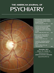
Looking Schizophrenia in the Eye
Schizophrenia is a complex and disturbing disorder. It is so common that nearly everyone knows someone with the condition, and it is so compelling that it has remained a subject of intense scientific scrutiny for over a century, pursued from every histopathological, molecular, social, and psychological perspective. The circles of investigation are both ever widening—encompassing genetics, infections, toxins, immigration, social status, and intergenerational exposures—and ever narrowing, as the focus now zooms in on specific domains of dysfunction, as seen from the brain and cognitive neuroscience, to regional anatomy and neurocircuitry, cell types, synapses, and molecules (1). Discoveries at each level may present a convincing narrative about the disease, but none has offered transformative interventions or cures.
Often a fascinating story line for schizophrenia begins and pauses, allegedly disproved by a different approach, or perhaps embodying a paradigm that transcends the dogma of the day. I recall meeting Dr. Janice Stevens as a medical student on a National Institute of Mental Health research elective and being enthralled by her knowledge and grasp of neurodegenerative theories of schizophrenia (2). I was later told that contradictory evidence demonstrating developmental origins for the disease had disproved her ideas. Nowadays, the developmental and degenerative views are incontrovertibly accepted, although they are seldom jointly considered. When novel findings are not replicated, the methodology is commonly implicated for having produced a false discovery, but the converse idea is seldom voiced. Only as the tools of our science advance do we rediscover previously known clues about the disease, including that early trauma and stress have a role in multiplying disease risk (3); that some heritability is associated with HLA (human leukocyte antigen) loci, as first shown in the earliest genetic studies (4) but now demonstrated by the most advanced genome-wide association studies (5); and that there are contributions to the illness from traumatic brain injury, as first described by Kraepelin from his observations of cases (6) but more securely accepted on the basis of a contemporary family study design (7). In schizophrenia, everything old is new again.
In 1928, for example, Jacobi and Winkler (8) demonstrated clear hydrocephalus in 18 of 19 chronic schizophrenia patients using pneumoencephalography, a technique in which air was introduced into the CSF through lumbar puncture and serial X-ray images were taken as the subject was rotated in space such that the air bubble could illuminate the cerebral ventricular contours. It took another half century and a technological revolution in neuroimaging for this seminal observation to gain the attention it deserved, when Johnstone and colleagues showed widened ventricles in schizophrenia using CT imaging in 1976 (9).
Now, in this issue of the Journal, Meier and colleagues (10) report a very intriguing finding for schizophrenia and psychosis using direct imaging of retinal microvessels in over 900 members of the Dunedin birth cohort at age 38. Probands with schizophrenia, and even those with only transient psychotic symptoms in childhood, had significantly wider venular calibers than other cohort members, including those with persistent depression. The dilated microvenules were unexplained by measurable confounders of illness, including antipsychotic treatment, or by several medical comorbidities frequently seen in persons with schizophrenia, including hypertension, diabetes, and tobacco dependence. Retinal imaging is an intriguing tool for understanding the etiopathophysiology of schizophrenia, but these findings using the newest tools for in vivo imaging of the vasculature to find a biomarker for schizophrenia have other implications as well.
In keeping with the theme of this editorial, this new finding builds on a long history of inquiry into the association between microvessel abnormalities and the risk for schizophrenia, as reviewed in this journal a half century ago (11), with the early studies using microscopic examinations of the nail fold capillary beds (12). The findings in these earlier studies suggest that the finding of widened retinal venules may not be specific to cerebral microvessels, but also be pertinent to other organ systems.
Notably, widened venules are particularly associated with systemic inflammatory conditions (13) as well as other schizophrenia-associated comorbidities, including cigarette smoking, high blood pressure, and obesity (14). Although endothelial dysfunction and inflammation are acknowledged to be associated with schizophrenia (15), this latest finding advances the field by confirming that the vascular finding (microvenule dilation) was even present in cohort members who only had some childhood psychotic symptoms and never developed mental illness. This suggests that the pathophysiology of the vascular abnormality is related to the initiating vulnerability for the disease itself and is not a consequence of the lifestyle related to the illness or of having received treatment for it. Based on the literature, the widened microvenules appear nonspecific and may also have multiple origins in schizophrenia. They could arise from prenatal and early-life infections or hypoxia, or from other adversities, such as susceptibility genes that interact with infections, stress, or toxic exposures, including cigarette smoking.
It is intriguing also to ponder whether decreased cerebral perfusion is a consequence of a microvascular dysfunction in addition to (or rather than) a consequence of intrinsic neural dysfunction. In a setting of venous dilation, sympathetic activity, which constricts arterioles, may leave too much blood in the venous system, decreasing venous return and reducing blood delivery to vital organs like the brain. Even if the neural vasculature is not subject to much influence by the autonomic nervous system (16), a blood-brain barrier disturbance may exist in schizophrenia that transmits this disturbance to the brain. Vascular dysfunction may explain or exacerbate reduced regional blood flow and even produce pathology in the brain, which may be exacerbated by stress and sympathetic discharges. Our knowledge of the etiology of wider venule caliber is nascent, but the studies linking hypoxia and past and current cigarette smoking, inflammation, and endothelial dysfunction to wider venules provide some clues.
The Meier et al. study in this issue reminds us that the vast proportion of genes and molecules associated with psychiatric conditions will also have other roles throughout the body. Dysfunction in these molecules or their regulation may produce systemic effects and not merely influence the brain and behavior. Likewise, vast numbers of the molecular mediators of venule caliber may have direct effects on brain and behavior that are independent manifestations of the molecular substrates or consequences of pathologically widened peripheral venules. Roles in both neurobiology and the dilation of microvenules are held by an array of players, including essential fatty acids (arachidonic acid or its derivatives), nitric oxide signaling, potassium and calcium ion channels, cellular energetic pathways, and numerous intracellular signaling cascades. Infections, stress, and illnesses are likewise systemic and not specific to brain function, such that the effector routes between numerous exposures and behavior could be conveyed via vascular alterations.
Schizophrenia and other serious psychiatric disorders gained resources and enhanced validity, as well as some reduction in stigma, when they were acknowledged to be brain disorders. Perhaps the next era in psychiatry must approach schizophrenia as a systemic disease that first presents as altered behavior rather than as a brain disease per se. We might feasibly learn more about the disease from its comorbidities than from enhanced measures of brain functioning. This step would be yet another echo from the idea that schizophrenia may be a form of familial diabetes (17).
1 : Toward the future of psychiatric diagnosis: the seven pillars of RDoC. BMC Med 2013; 11:126Crossref, Medline, Google Scholar
2 : Anatomy of schizophrenia revisited. Schizophr Bull 1997; 23:373–383Crossref, Medline, Google Scholar
3 : Communication deviance in parents of families with adoptees at a high or low risk of schizophrenia-spectrum disorders and its associations with attributes of the adoptee and the adoptive parents. Psychiatry Res 2011; 185:66–71Crossref, Medline, Google Scholar
4 : HLA antigens and schizophrenia. Lancet 1980; 1:765Crossref, Medline, Google Scholar
5
6 : Dementia Praecox and Paraphrenia. Translated by Barclay RM; edited by Robertson GM. Edinburgh, E & S Livingstone, 1919Google Scholar
7 : Traumatic brain injury and schizophrenia in members of schizophrenia and bipolar disorder pedigrees. Am J Psychiatry 2001; 158:440–446Link, Google Scholar
8 : Encephalographische Studien an Schizophrenen. Arch Psychiatrie 1928; 84:208–226Crossref, Google Scholar
9 : Cerebral ventricular size and cognitive impairment in chronic schizophrenia. Lancet 1976; 2:924–926Crossref, Medline, Google Scholar
10 : Microvascular abnormality in schizophrenia as shown by retinal imaging. Am J Psychiatry 2013; 170:1451–1459Link, Google Scholar
11 : Psychological correlates of capillary morphology in schizophrenia. Am J Psychiatry 1965; 122:444–446Link, Google Scholar
12 : Capillary morphology of the nailfold in the mentally ill. J Neuropsychiatr 1964; 5:225–234Medline, Google Scholar
13 : Retinal vascular caliber, cardiovascular risk factors, and inflammation: the Multi-Ethnic Study of Atherosclerosis (MESA). Invest Ophthalmol Vis Sci 2006; 47:2341–2350Crossref, Medline, Google Scholar
14 : Relative importance of systemic determinants of retinal arteriolar and venular caliber: the Atherosclerosis Risk in Communities Study. Arch Ophthalmol 2008; 126:1404–1410Crossref, Medline, Google Scholar
15 : Theories of schizophrenia: a genetic-inflammatory-vascular synthesis. BMC Med Genet 2005; 6:7Crossref, Medline, Google Scholar
16 : The vascular conducted response in cerebral blood flow regulation. J Cereb Blood Flow Metab 2013; 33:649–656Crossref, Medline, Google Scholar
17 : Family history of type 2 diabetes in schizophrenic patients. Lancet 1989; 1:495Crossref, Medline, Google Scholar


