In Search of the Emotional Self: An fMRI Study Using Positive and Negative Emotional Words
Abstract
OBJECTIVE: The authors used functional magnetic resonance imaging (fMRI) to define the neural regions mediating self-referential processing of emotional stimuli and to explore how these regions are influenced by the emotional valence of the stimulus. METHOD: Ten healthy subjects were presented with words describing positive and negative personality traits during fMRI scanning in three different conditions. In the self-referential processing condition, subjects judged whether they thought each trait described them. In the other-referential processing condition, subjects judged whether the stimulus described a generally desirable trait. In the letter-recognition control condition, subjects indicated whether the word contained a specific target letter. RESULTS: The self-referential condition induced bilateral activation in the dorsomedial prefrontal cortex, whereas the other-referential condition induced activation in lateral prefrontal areas. Activation in the right dorsomedial prefrontal cortex was unique to the self-referential condition regardless of the valence of the words, although positive words produced a more robust activation than did negative words. In the self-referential condition, differences between the processing of positive and negative words were seen in regions outside the medial frontal cortex, with reductions in the insula, temporal and occipital regions, and inferior parietal regions associated with negative words. CONCLUSIONS: A widely distributed network of brain areas contributes to emotional processing. Among these regions, the right dorsomedial prefrontal cortex is one main area mediating self-reference. By providing a personal perspective in the evaluation of emotional stimuli, the right dorsomedial prefrontal cortex may mediate cognitive processes, such as those involved in psychotherapy, that guide self-regulation of emotional experience.
Cognitive models of emotion suggest that mood disturbances, such as those seen in depression, may reflect the exaggeration of normal emotional responses or abnormalities in emotional processing (1). As suggested by these models, imaging studies of the brain circuitry underlying normal emotional behaviors may contribute to a better understanding of the neural basis of affective disorders.
Several brain imaging studies have previously linked components of emotional behaviors to limbic and paralimbic structures (insula, amygdala, medial temporal cortex, anterior cingulate) and to subcortical regions (thalamus, caudate, hypothalamus), as well as to medial, dorsolateral, and orbitofrontal regions (2–4). An unresolved question is whether the same neural structures mediate both positive and negative emotions. Some studies have supported the hypothesis that positive and negative emotions are processed in different neural structures (2, 3). Davidson and Irwin (5) proposed that the right prefrontal cortex is more active during negative emotions such as fear, disgust, or sadness than during positive emotions. Other authors have suggested a nonlateralized prefrontal pattern in negative and positive emotion in which ventral areas are active during negative emotion and dorsal areas are active during positive emotion (6, 7). In addition, while some studies (8, 9) have reported that the anterior cingulate and medial prefrontal cortex play a specific role in the cognitive processing of emotional stimuli, little is known about the neural basis of self-referential processing of these types of stimuli.
Recent neuroimaging studies of normal subjects, however, have suggested that the medial prefrontal cortex may be specifically involved in tasks requiring the processing of information relevant to the self (10, 11). Most of these studies have assessed self-referential processing by using cognitive tasks without emotional components. The role of medial prefrontal cortex regions in self-referential processing within an inherently emotional context remains unclear, although critically important to understanding inappropriate self-referencing in patients with depression.
The aim of the current study was to identify the brain regions mediating interactions between self-related processing and emotional processing. The experiment was designed to build on previous work demonstrating that words processed with reference to the self (some studies used nouns, not all of which had emotional valence) are generally better remembered than material processed in semantic terms (12). Numerous studies have established the reliability and validity of this phenomenon, labeled the “self-reference effect” (12). Paradigms that use emotional words to examine the self-referential effect allow assessment of self-related processes within an emotional context. Craik et al. (13), using a self-reference effect paradigm and positron emission tomography (PET), determined that self-encoding of adjectives describing personality traits yielded specific activation of the right dorsomedial prefrontal cortex. However, this study did not allow for comparison between stimuli with positive and negative emotional valences and therefore did not provide a basis for investigating the emotional bias toward negative stimuli in depression.
The current study used a self-reference effect paradigm with an event-related fMRI design to evaluate the modulation by valence of the neural response associated with self-evaluation of emotional words. Based on our previous PET scan study (13), we hypothesized that the dorsomedial prefrontal cortex, especially in the right hemisphere, would contribute to the integration of emotion and self-referential processing. Lateralized or nonlateralized valence effects were not additionally hypothesized, given the many inconsistencies in the published literature.
Method
Subjects
Fourteen healthy subjects (eight women, six men; mean age=26.4 years, SD=4.5, range=20–36), screened for absence of psychiatric and neurological disorders with a clinical interview, were recruited from the University of Toronto community. At the time of initial screening, participants’ mood was assessed with the Beck Depression Inventory (14). All subjects were fluent in English, were right-handed as judged by the Edinburgh Handedness Inventory (15), and had normal or corrected-to-normal visual acuity. After a complete description of the study to the subjects, written informed consent was obtained. The study was approved by research ethics boards at Baycrest Centre, Sunnybrook and Women’s College Health Sciences Centre, and the University of Toronto. Subjects were reimbursed for their participation and time. Four subjects whose fMRI time series had perceptible, residual head movements greater than 1 mm were excluded from further analysis. The remaining 10 subjects (seven women, three men; mean age=25.8 years, SD=4.4; mean number of years of education=17.9, SD=2.0; mean Beck Depression Inventory score=2.4, SD=2.9) were included in the study.
Verbal Stimuli
Thirteen lists of 10 personality-trait adjectives were constructed from Anderson’s list of personality-trait words (16). The lists were used in the judgment tasks described in the next section (additional lists were used in a subsequent recognition test that will be detailed elsewhere, in a separate report). Within each of the 13 lists, half of the words were unambiguously positive and half were negative, selected from the top 20% and bottom 20%, respectively, of Anderson’s sample. One list was used for practice trials. The other 12 lists were shown to the participants during the scan acquisitions. The study included three different judgment conditions: self-referential processing, other-referential processing, and letter recognition (used as the baseline control task). Four lists were assigned to each of three judgment conditions. Each positive and negative word occurred in only one of the lists, and each word was randomly assigned to only one of the three judgment conditions for each subject. Therefore, each subject received a unique protocol.
Task Design
The fMRI scans were acquired while patients performed the judgment tasks. Stimuli were generated by a personal computer with SuperLab Pro software (Cedrus Corp., San Pedro, Calif.) and appeared on a back-projection screen mounted outside the scanner bore; subjects viewed the screen using angled mirrors.
Depending on instructions presented before the start of each set, participants made one of three different judgments about the presented words. Subjects were not required to directly rate the valence itself. In the self-referential condition, subjects judged whether each trait described them. In the other-referential condition, participants judged whether the trait was socially desirable. In the letter-recognition control condition, participants judged whether the word contained a specific target letter (i.e., “e” or “a”; a different letter was used for each list). For all conditions, each word prompted a “yes” or “no” response, and subjects pressed a button with the right index or middle finger, respectively, to indicate their decision. Button presses were recorded by using an MRI-compatible keypad (Lumitouch, Lightwave Technologies, Richmond, B.C., Canada). Participants were not explicitly asked to remember the words.
Subjects performed each judgment condition four times across three scanning runs. At the onset of each condition, an instruction cue was presented for 9 seconds (e.g., “task: self condition”). Each condition contained trials consisting of two word types: negative words and positive words. Negative and positive words were randomly intermixed and separated by a fixation crosshair displayed on the center of the screen. Item order was counterbalanced such that positive and negative items followed each other equally often. Each list of adjectives constituted 10 trials. Each trial consisted of a fixation crosshair displayed for 500 msec followed by an adjective displayed for 4500 msec, then another fixation point displayed for 5000 msec. Participants responded by pressing a keypad at any time between the onset of the stimulus and the onset of the 500-msec fixation of the next trial. To control for order effects, blocks within a run (40 words per run, 10 words per block; a total of four blocks per run) were presented in random order, with no two consecutive blocks in the same condition. Both response and reaction times were recorded by SuperLab.
Encoding tasks were immediately followed by an unexpected recognition task. For the recognition task, subjects were instructed to discriminate between studied (old) words and unstudied (new) words. Each word prompted a “yes” (old) or “no” (new) response, and subjects pressed a button with the right index or middle finger, respectively, to indicate their decision.
Functional Imaging
Imaging was performed at Sunnybrook and Women’s College Health Sciences Centre by using a 1.5-T Signa MRI system (CV/i hardware, LX8.3 software; General Electric Medical Systems, Waukesha, Wis.) with a standard quadrature birdcage head coil. Structural magnetic resonance images were acquired by using a three-dimensional T1-weighted spoiled gradient recall acquisition sequence (TR=12.4 msec, TE=5.4 msec, flip angle 35°, 22×16.5 field of view, 256×196 acquisition matrix, 124 axial slices, 1.4 mm thickness). Functional images were collected by using a T2*-weighted pulse sequence with spiral k-space readout, offline gridding, and reconstruction (TE=40 msec, TR=2000 msec, flip angle=80°, 90×90 effective acquisition matrix, 20 cm field of view, 24 axial slices, 5 mm thickness) optimized for sensitivity to blood-oxygenation-level-dependent (BOLD) signal contrast (17). Functional data were acquired in three separate runs, each lasting 7 minutes and 56 seconds (including an initial 40 seconds for machine equilibration).
Statistical Analysis
Analysis of fMRI data
Images of brain activation were computed and overlaid on anatomic images by using the Analysis of Functional NeuroImaging (AFNI) program (18). Time series data were first spatially coregistered to correct for head motion by using a three-dimensional Fourier transform interpolation and were then detrended to a constant reference scan by using a fifth-order polynomial. The data were subjected to two analyses: one to examine the effects of self-processing regardless of word valence, and the other to clarify the role of valence.
Analysis of hemodynamic response
Percent changes in signal intensity during the self-referential and other-referential conditions with respect to the letter-recognition control condition were analyzed by using voxelwise correlations of the self-referential and other-referential time series with square-wave reference vectors (19) shifted to account for the delay in hemodynamic response. This method produced two activation images per participant: one for the self-referential condition versus the letter-recognition control condition, and one for the other-referential condition versus the letter-recognition control condition. These activation images were then transformed into Talairach coordinates (18, 20) and smoothed by using a Gaussian filter with a 6-mm full width at half maximum to increase the signal-to-noise ratio. The latter step was performed to facilitate the subsequent group analysis consisting of a mixed-effect, voxelwise two-factor analysis of variance (ANOVA) of the percent changes in signal intensity, with judgment condition (self- or other-referential) as the within-subjects factor and subject as a random factor. Because of our a priori hypothesis targeting the medial prefrontal region, the statistical cutoff for the main comparison between the self and other conditions was set at p<0.001, uncorrected.
Analysis of valence effect
In the second analysis, we contrasted positive and negative words to determine the effect of valence in self-referential processing. To this end, we compared responses to positive and negative words in the self-referential condition relative to the letter-recognition control condition. AFNI was used to deconvolve the hemodynamic response function on a voxelwise basis from the time series data to interpret the activations associated with positive and negative words randomly presented within a given block. The best linear least-squares fit was calculated for the following model parameters: constant baseline, linear trend in time series, and BOLD response deviation from baseline for each condition (self-referential processing of positive words and self-referential processing of negative words). This fit of the parameters produced an estimate of the hemodynamic response for the image 0–5 TR (10 seconds) after the stimulus onset for each condition relative to the control condition. Subsequently, activation images were created corresponding to 3 TR after the stimulus onset for each of the conditions contrasted with the corresponding letter-recognition condition (self-referential processing of positive words versus letter recognition for positive words, self-referential processing of negative words versus letter recognition for negative words). The images were then transformed into Talairach coordinates and spatially smoothed as reported earlier.
The subsequent group analysis consisted of a mixed-effect, voxelwise two-factor ANOVA with condition (self-referential processing of positive words, self-referential processing of negative words) as the within-subject factor and subject as a random factor. The statistical cutoff for the main comparison of self-referential processing of positive and negative words was set at p<0.001, uncorrected. Post hoc specific contrast manipulations were then investigated in more detail.
In addition, because both the self-referential and the letter-recognition conditions involved both explicit and implicit processing of the emotional aspects of the words, it was also important to determine whether the comparison of self-referential processing with letter recognition removed a specific valence effect. This possibility was assessed by contrasting activation associated with positive and negative words across all the different conditions.
Results
Behavioral Results
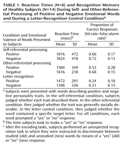
Table 1 shows the mean reaction times subjects took to make judgments for each word condition and valence type. Due to technical problems, response times were recorded for only six subjects. A repeated measures ANOVA with judgment condition and valence as the within-subject factors showed that neither the main effect of judgment condition (F=1.87, df=2, 4, n.s.) nor of valence (F=0.07, df=1, 5, n.s.) was significant. Likewise, the judgment-by-valence interaction was not statistically significant (F=0.09, df=2, 4, n.s.).
Details of the recognition data will be the subject of a separate report. In short, recognition was significantly higher in the self- and other-referential conditions than in the letter-recognition condition, and the results suggested an effect of self-reference on memory performance.
fMRI Results
Analysis without valence
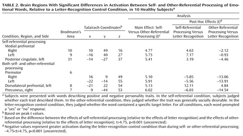
Brain regions with activations associated with self- and other-referential processing, irrespective of word valence, are listed in Table 2. As reported in our previous PET study (13) and consistent with our hypothesis, the self-referential condition was associated with left and right dorsomedial prefrontal cortex activations (Figure 1). In addition, this analysis showed signal activation unique to the self-referential condition in the left posterior cingulate (Brodmann’s area 31), which has not been previously identified. Processing emotional words in both the self- and other-referential conditions induced BOLD signal activations in the lateral prefrontal areas and reductions in the parietal and premotor regions (Table 2). No regions showed significant neural activations specific to the other-referential condition.
Valence effect
To address the possibility that there might be an effect of valence within self-referential processing, we compared processing of positive and negative words in the self-referential condition. A significant difference between the two valence conditions in the area of primary interest, the right dorsomedial prefrontal cortex, was not found (Table 3). However, post hoc analyses showed that both the right and left dorsomedial prefrontal cortex were active during self-referential processing of both positive and negative words, with positive words producing a more robust activation than did negative words (Table 3). Taken together these results suggest that the dorsomedial prefrontal cortex, especially on the right, is one main area for self-referential processing and is not independently modulated by positive or negative valence.
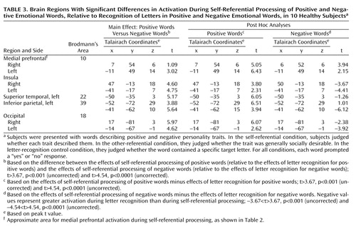
Although the comparison of self-referential processing of positive and negative words did not show significant differences in activation in the dorsomedial prefrontal cortex, the comparison did reveal significant activations for positive words in the left and right insula and in the temporal, occipital, and inferior parietal regions (Table 3). No regions showed significant neural activations specific to the processing of negative words.
Post hoc analyses revealed that the main effect in the comparison of self-referential processing of positive and negative words was due not to increases with positive words but rather to significant reductions during the processing of negative words in the ventral and lateral paralimbic regions—the left and right insula—and more posterior regions, including the left inferior parietal and the right and left occipital cortex (Figure 2 and Table 3). Finally, examination of activations associated with positive and negative words independent of task (i.e., combining results from the self- and other-referential conditions and the letter-recognition control condition, then comparing effects for positive versus negative words) revealed no significant valence differences.
Discussion
The goal of this study was to identify brain regions mediating self-referential processing of positive and negative emotional words. The main finding was that emotional stimuli processed in a personal context were associated with a different pattern of brain activations than were identical stimuli considered in a general context. Self-referential processing of emotional words induced a unique activation in the dorsomedial prefrontal cortex. Moreover, the activation seen in the dorsomedial prefrontal cortex, especially on the right, was present irrespective of the valence of the words, suggesting that this region is involved in self-referential processing of all emotional stimuli, regardless of emotional valence.
The fMRI results additionally showed that the magnitude of the signal in the self-referential condition was greater than in the other-referential condition in left dorsomedial prefrontal cortex regions (Brodmann’s area 8/9), consistent with our earlier study (13) and a large literature on semantic processing of words and the hemispheric encoding retrieval asymmetry (HERA) model (21). Likewise, the association of self-reference with posterior cingulate activity replicates earlier findings implicating the posterior cingulate in aspects of emotional processing (22).
One possible explanation for the selective right dorsomedial prefrontal cortex activity might be the targeted use of strictly emotional words. However, processing of emotional words is not consistently associated with activations of the dorsomedial prefrontal cortex across brain imaging studies. One PET study (23) found increased activity in the medial prefrontal regions when subjects passively viewed emotional words relative to passively viewing animal names, while another study (24) showed brain activation mainly in the retrosplenial cortex associated with threat-related words and no activation in the dorsomedial prefrontal cortex.
We suggest that the dorsomedial prefrontal cortex activity is not likely to be related to emotional content per se but is due to the self-referential processing of the emotional stimuli. Several converging lines of evidence support this interpretation. The right dorsomedial prefrontal cortex has been reliably activated across a wide range of emotional tasks with or without verbal materials, such as recollection of personally affect-laden life events (25), attention to subjective feeling (8), and processing of emotion-related meanings (7). All these tasks can be characterized as requiring access to, or manipulation of, explicit representation of different aspects of the self and integration of these aspects with emotional reactions and experience.
The new finding from this study is that the right dorsomedial prefrontal cortex is activated during self-evaluation regardless of whether the valence of the words is positive or negative. Unlike other works showing hemispheric asymmetry in the mediation of positive and negative emotions (5), we found no evidence for laterality effects due to the valence of the emotional words.
A further finding is the difference in regions outside of the prefrontal cortex that were identified with respect to the self-referential processing of positive and negative emotional words. The self-referential processing of negative words induced significant BOLD signal reduction in the right inferior parietal lobe, the left and right insula, and more posterior regions. One interpretation is that the processing of negative words in the self-referential condition necessitated more cognitive effort than the processing of positive words. This interpretation would be consistent with recent suggestions by Pochon et al. (26) of a negative modulation of limbic activity by task complexity.
The insula and parietal regions are also associated with anxiety, depression, and provocation of feelings of sadness (27), suggesting that these regions are more generally involved in the integration of internal states. The present experiment was not designed to elicit explicit emotional feeling states in subjects. However, to avoid feeling emotions and maintain a self-protective mode, subjects may have attempted to inhibit their emotional responses during self-processing of negative words (28).
The dorsomedial prefrontal cortex and the adjacent anterior cingulate have direct reciprocal connections with the inferior parietal cortex, dorsolateral prefrontal cortex, and posterior cingulate. The dorsomedial prefrontal cortex and the dorsal anterior cingulate also communicate with the paralimbic structures, the ventromedial prefrontal cortex, and brainstem regions indirectly through the rostral and subgenual cingulate areas (29). Therefore, by virtue of the capacity to receive inputs from these limbic and paralimbic regions and to project to other prefrontal areas, the dorsomedial prefrontal cortex is a suitable region for integrating cognitive processing with emotional reactions and experience.
The human “self model” is a theoretical construct comprising essential features such as feelings of continuity and unity, experience of agency, and experience of a body-centered perspective (30). We propose that one specific role of the right dorsomedial prefrontal cortex is to represent states of an emotional episodic “self” and then to process emotional stimuli with a personally relevant perspective. This proposition is in line with studies showing activations within both the left and right dorsomedial prefrontal cortex during “theory of mind” tasks (31). Because emotions generally signal issues related to the self, subjects may use emotional cues during some theory of mind tasks to differentiate self from other; this self-related emotional processing is indicated by an increase of activity in the right dorsomedial prefrontal cortex.
This study had some limitations that must be considered in interpreting the findings. Owing to technical difficulties, only 10 of the original 14 subjects were included in the final analysis. Thus, the study group size may have been underpowered to detect a valence effect. Further studies with larger numbers of subjects are therefore needed to confirm the results.
To assess self-processing within an emotional context, we used an incidental encoding task in which subjects made different evaluative judgments about emotional words. Behavioral results showed that response times for judging each word were similar in the self- and other-referential conditions. However, data on response reaction times were available for only six subjects. Therefore, we cannot definitely rule out the possibility that, because of the small number of subjects, the differences in reaction times may actually reflect real differences in brain activation between the self- and other-referential conditions.
Positive and negative emotional words were not matched for the arousal associated with them. Negative stimuli are typically more arousing than positive words, and fMRI differences between the word types may be due to this potential confound. However, we found that positive words induced greater activation than negative words. Moreover, self-referential processing of negative words was associated with BOLD reductions in several regions, an unlikely effect of emotional arousal, as suggested by past observations (32).
To replicate our previous PET findings (13), we used the same paradigm with emotional words for each condition. Further studies including neutral words may help to confirm our findings on the role of the dorsomedial prefrontal cortex in self-referential processing of emotional stimuli with different valences.
In summary, our findings provide support for the involvement of a widely distributed set of brain regions in the processing of self-referential information. Among these regions, the right dorsomedial prefrontal cortex appears most specific for the more subjective, perspective-taking aspects involved in emotional evaluation. The common role of the dorsomedial prefrontal cortex in evaluating both positive and negative stimuli further suggests that the dysfunction of the dorsomedial prefrontal cortex frequently reported in mood disorders (33) may subserve the bias of emotional processing in depression.
 |
 |
 |
Presented in part at the seventh annual meeting of the Organization for Human Brain Mapping, Brighton, U.K., June 10–14, 2001. Received Aug. 6, 2002; revision received March 17, 2003; accepted March 26, 2003. From the Rotman Research Institute, Baycrest Centre for Geriatric Care, Toronto; the Department of Psychiatry and CNRS UMR 7593, Salpétrière Hospital; and the Departments of Medical Biophysics, Psychology, and Psychiatry, University of Toronto, Toronto. Address reprint requests to Dr. Fossati, Department of Psychiatry, CNRS UMR 7593, Salpétrière Hospital, 47 boulevard de l’hôpital, 75651 Paris Cedex 13, France; [email protected] (e-mail). Supported by the Sandra Rotman Program in Neuropsychiatry (Dr. Mayberg) and a grant from the Fondation Singer Polignac and Société de Secours des Amis des Sciences (Dr. Fossati). The authors thank Rhonda Walcarius for technical support and J.B. Pochon for comments on a previous version of the manuscript.

Figure 1. Areas in the Dorsomedial Prefrontal Cortex Showing Significant Increase in Activation During Self-Referential Processing of Emotional Words, Relative to Other-Referential Processing, in 10 Healthy Subjectsa
aSubjects were presented with words describing positive and negative personality traits during fMRI scanning. In the self-referential condition, subjects judged whether they thought each trait described them. In the other-referential condition, subjects judged whether the trait was generally socially desirable. Areas highlighted in red-yellow show contiguous voxels exceeding the statistical cutoff (–4.75<t>4.75, p<0.001, uncorrected). The y coordinate indicates the location of the coronal slice.

Figure 2. Areas in the Insula Showing Significant Increase in Activation During Self-Referential Processing of Positive and Negative Emotional Words, Relative to Recognition of Letters in Positive and Negative Emotional Words, in 10 Healthy Subjectsa
aSubjects were presented with words describing positive and negative personality traits during fMRI scanning. In the self-referential condition, subjects judged whether they thought each trait described them. In the letter-recognition condition, subjects indicated whether the word contained a specific target letter. In the left panel, red-yellow highlighting shows contiguous voxels exceeding the statistical cutoff (–4.54<t>4.54, p<0.001, uncorrected) and indicates areas with greater activation during self-referential processing of positive words than during self-referential processing of negative words, relative to the letter-recognition condition. In the right panel, blue highlighting shows contiguous voxels exceedingthe statistical cutoff (–4.54<t>4.54, p<0.001, uncorrected) and indicates areas with decreased activation during self-referential processing of negative words, relative to the letter-recognition control condition. The y coordinates indicate the locations of the coronal slices.
1. Williams JMG, Watts FN, MacLeod C, Mathews A: Cognitive Psychology and Emotional Disorders. Chichester, UK, John Wiley & Sons, 1988Google Scholar
2. George MS, Ketter TA, Parekh PI, Horwitz B, Herscovitch P, Post RM: Brain activity during transient sadness and happiness in healthy women. Am J Psychiatry 1995; 152:341–351Link, Google Scholar
3. Lane RD, Reiman EM, Ahern GL, Schwartz GE, Davidson RJ: Neuroanatomical correlates of happiness, sadness, and disgust. Am J Psychiatry 1997; 154:926–933Link, Google Scholar
4. Morris JS, Frith CD, Perrett DI, Rowland D, Young AW, Calder AJ, Dolan RJA: A differential neural response in the human amygdala to fearful and happy facial expressions. Nature 1996; 383:812–815Crossref, Medline, Google Scholar
5. Davidson RJ, Irwin W: The functional neuroanatomy of emotion and affective style. Trends Cogn Sci 1999; 3:11–21Crossref, Medline, Google Scholar
6. Schneider F, Gur RE, Mozley L, Smith RJ, Mozley P, Censits DM, Alavi A, Gur R: Mood effects on limbic blood flow correlate with emotional self-rating: a PET study with oxygen-15 labeled water. Psychiatry Res 1995; 61:265–283Crossref, Medline, Google Scholar
7. Teasdale JD, Howard RJ, Cox SG, Ha Y, Brammer MJ, Williams SCR, Checkley SA: Functional MRI study of the cognitive generation of affect. Am J Psychiatry 1999; 156:209–215Abstract, Google Scholar
8. Lane RD, Fink GR, Chau PM, Dolan RJ: Neural activation during selective attention to subjective emotional responses. Neuroreport 1997; 22:3969–3972Crossref, Google Scholar
9. Lane RD, Reiman EM, Axelrod B, Yun LS, Holmes A, Schwartz GE: Neural correlates of levels of emotional awareness: evidence of an interaction between emotion and attention in the anterior cingulate cortex. J Cogn Neurosci 1998; 10:525–535Crossref, Medline, Google Scholar
10. Gusnard DA, Akbudak E, Shulman GL, Raichle ME: Medial prefrontal cortex and self-referential mental activity: relation to a default mode of brain function. Proc Natl Acad Sci USA 2001; 98:4259–4264Crossref, Medline, Google Scholar
11. Johnson SC, Baxter LC, Wilder LS, Pipe JG, Heiserman JE, Prigatano GP: Neural correlates of self-reflection. Brain 2002; 125:1808–1814Crossref, Medline, Google Scholar
12. Symons CS, Johnson BT: The self-reference effect in memory: a meta-analysis. Psychol Bull 1997; 121:371–394Crossref, Medline, Google Scholar
13. Craik FIM, Moroz TM, Moscovitch M, Stuss DT, Winokur G, Tulving E, Kapur S: In search of the self: a positron emission tomography investigation. Psychol Sci 1999; 10:26–34Crossref, Google Scholar
14. Beck AT, Ward CH, Mendelson M, Mock J, Erbaugh J: An inventory for measuring depression. Arch Gen Psychiatry 1961; 4:561–571Crossref, Medline, Google Scholar
15. Williams SM: Factor analysis of the Edinburgh Handedness Inventory. Cortex 1986; 22:325–326Crossref, Medline, Google Scholar
16. Anderson N: Likableness ratings of 555 personality-trait words. J Pers Soc Psychol 1968; 9:272–279Crossref, Medline, Google Scholar
17. Glover GH, Lai S: Self-navigated spiral fMRI: interleaved versus single-shot. Magn Reson Med 1998; 39:361–368Crossref, Medline, Google Scholar
18. Cox RW: AFNI: software for analysis and visualization of functional magnetic resonance neuroimages. Comput Biomed Res 1996; 29:162–173Crossref, Medline, Google Scholar
19. Bandettini PA, Jesmanowicz A, Wong EC, Hyde JS: Processing strategies for time-course data sets in functional MRI of the human brain. Magn Reson Med 1993; 30:161–173Crossref, Medline, Google Scholar
20. Talairach J, Tournoux P: Co-Planar Stereotaxic Atlas of the Human Brain: Three-Dimensional Proportional System. New York, Thieme Medical, 1988Google Scholar
21. Tulving E, Kapur S, Craik FIM, Moscovitch M, Houle S: Hemispheric encoding/retrieval asymmetry in episodic memory: positron emission tomography findings. Proc Natl Acad Sci USA 1994; 91:2016–2020Crossref, Medline, Google Scholar
22. Maddock RJ: The retrosplenial cortex and emotion: new insights from functional neuroimaging of the human brain. Trends Neurosci 1999; 22:310–316Crossref, Medline, Google Scholar
23. Beauregard M, Chertkow H, Bub D, Murtha S, Dixon R, Evans A: The neural substrate for concrete, abstract and emotional words lexica: a positron emission tomography study. J Cogn Neurosci 1997; 9:441–473Crossref, Medline, Google Scholar
24. Maddock RJ, Buonocore MH: Activation of left posterior cingulate gyrus by the auditory presentation of threat-related words: an fMRI study. Psychiatry Res 1997; 75:1–14Crossref, Medline, Google Scholar
25. Reiman EM, Lane RD, Ahern GL, Schwartz GE, Davidson RJ, Friston KJ, Yun L-S, Chen K: Neuroanatomical correlates of externally and internally generated human emotion. Am J Psychiatry 1997; 154:918–925Link, Google Scholar
26. Pochon JB, Levy R, Fossati P, Lehericy S, Poline JB, Pillon B, Le Bihan D, Dubois B: The neural system that bridges reward and cognition in humans: an fMRI study. Proc Natl Acad Sci USA 2002; 99:5669–5674Crossref, Medline, Google Scholar
27. Mayberg HS, Liotti M, Brannan SK, McGinnis S, Mahurin RK, Jerabek PA, Silva JA, Tekell JL, Martin CC, Lancaster JL, Fox PT: Reciprocal limbic-cortical function and negative mood: converging PET findings in depression and normal sadness. Am J Psychiatry 1999; 156:675–682Abstract, Google Scholar
28. Beauregard M, Levesque J, Bourgouin P: Neural correlates of conscious self-regulation of emotion. J Neurosci 2001; 21:RC165Google Scholar
29. Chavis DA, Pandya DN: Further observations on corticofrontal connections in the rhesus monkey. Brain Res 1976; 117:369–386Crossref, Medline, Google Scholar
30. Gallagher S: Philosophical conceptions of the self: implications for cognitive science. Trends Cogn Sci 2000; 4:14–21Crossref, Medline, Google Scholar
31. Castelli F, Happé F, Frith U, Frith CD: Movement and mind: a functional imaging study of perception and interpretation of complex intentional movement patterns. Neuroimage 2000; 12:314–325Crossref, Medline, Google Scholar
32. Critchley HD, Corfield DR, Chandler MP, Mathias CJ, Dolan RJ: Cerebral correlates of autonomic cardiovascular arousal: a functional neuroimaging investigation in humans. J Physiol 2000; 523:250–270Crossref, Google Scholar
33. Mayberg HS: Modulating dysfunctional limbic-cortical circuits in depression: towards development of brain-based algorithms for diagnosis and optimised treatment. Br Med Bull 2003; 65:193–207Crossref, Medline, Google Scholar