Higher Expression of Serotonin 5-HT2A Receptors in the Postmortem Brains of Teenage Suicide Victims
Abstract
OBJECTIVE: Abnormalities of serotonin (5-HT) receptor subtypes have been observed in the postmortem brains of adult suicide victims; however, their role in teenage suicide is unexplored. The authors examined whether 5-HT2A receptor subtypes are altered in the postmortem brains of teenage suicide victims. METHOD: Levels of 5-HT2A receptors were determined through examination of [125I] LSD binding, protein expression (by use of Western blotting with a specific 5-HT2A receptor antibody), and mRNA (by means of quantitative reverse transcription polymerase chain reaction) in the prefrontal cortex, hippocampus, and nucleus accumbens of 15 teenage suicide victims and 15 normal matched teenage subjects. The cellular localization of the 5-HT2A receptors was determined by means of gold immunolabeling. RESULTS: The authors observed significantly higher [125I]LSD binding in the prefrontal cortex and greater protein expression and mRNA levels in the prefrontal cortex and hippocampus but not in the nucleus accumbens of suicide victims, compared with normal subjects. Greater protein expression was localized on pyramidal cells in cortical layer V but not in other cortical layers or in the surrounding neuropil of the prefrontal cortex of teenage suicide victims. CONCLUSIONS: The evidence indicates higher levels of 5-HT2A receptor, protein, and mRNA expression in the prefrontal cortex and hippocampus, which have been implicated in emotion, stress, and cognition. There was no higher level in the nucleus accumbens, which has been implicated in drug dependence and craving. Our findings suggest that a higher level of 5-HT2A receptors may be one of the neurobiological abnormalities associated with teenage suicide.
Suicide is a major public health concern (1) and the ninth leading cause of death; about 30,000 individuals die by suicide each year in the United States alone (2). In the young, it is the second most frequent cause of death. Suicidal behavior is often associated with mental disorders, such as depression, bipolar illness, and personality disorders. Besides psychiatric illnesses, other risk factors include a family history of suicide, a family history of psychiatric disorders, psychosocial stressors, impulsivity, and aggression (3). Abnormalities in neurobiological mechanisms may be another risk factor for suicide. Several studies (4, 5) have suggested that the major driving factor leading to suicide in teenagers is aggressive and impulsive behavior. Since abnormalities in serotonin (5-HT) function have been implicated in impulsive/aggressive behaviors (6), it is possible that the pathophysiology of teenage suicide may be associated with abnormal serotonin function.
Studies of postmortem brain samples from adult suicide victims have suggested that abnormalities of 5-HT receptor subtypes are associated with suicide (for a review, see Gross-Isseroff et al. [7]). In particular, it has been reported by several investigators, although not confirmed by all, that one of the serotonin receptor subtypes, the 5-HT2A receptor, is found in higher than normal levels in the postmortem brains of adult suicide victims (7–10). Further support for this concept comes from studies that indicate a greater number of 5-HT2A receptors in the platelets of suicidal patients with different mental disorders; this higher level appears to be independent of diagnosis (11, 12). On the basis of these findings, we previously proposed that platelet 5-HT2A receptors might serve as a biological marker of suicidal behavior (11).
Although serotonergic abnormalities and 5-HT2A receptors have been studied in the brains of adult suicide victims, to our knowledge, serotonin receptor subtypes, specifically 5-HT2A receptors, have not been studied in the postmortem brains of teenage suicide victims. In addition, earlier studies of 5-HT2A receptors in the postmortem brains of adult suicide victims showed higher 5-HT2A receptor numbers on the basis of the greater binding of 5-HT2A receptor ligands to their binding sites. Thus, whether higher 5-HT2A receptor numbers in suicide victims are the result of more active binding sites or are associated with abnormalities at the level of transcription and/or translation of 5-HT2A receptors is not yet known.
Therefore, in the present investigation, we examined the association of 5-HT2A receptors and teenage suicide, not only by examining the binding characteristics of 5-HT2A receptors (to replicate the findings in adult suicide victims), but also by studying their gene transcription and translation in the prefrontal cortex, hippocampus, and nucleus accumbens in teenage suicide victims and nonsuicide subjects. Furthermore, we localized the changes in 5-HT2A receptors at the cellular level in various cortical layers by using gold immunolabeling.
Method
5-HT2A receptor levels were determined in the prefrontal cortex (Brodmann’s areas 8/9), hippocampus, and nucleus accumbens (all from the right hemisphere) in brains obtained from 15 teenage suicide victims and 15 psychiatrically normal teenage subjects who died of other causes, herein referred to as normal subjects. Postmortem brain tissue was obtained from the Brain Collection Program at the Maryland Psychiatric Research Center in Baltimore, Md., in collaboration with the Medical Examiner’s Office of the State of Maryland. Brain samples were free of neuropathological abnormalities and HIV antibodies. Toxicological data were obtained by analysis of urine and blood samples.
Diagnostic Methods
All study victims were diagnosed by means of the Structured Clinical Interview for DSM-III-R (13) and the Diagnostic Evaluation After Death (14); diagnoses were based on the information obtained from interviewing at least one family member and from all available medical records. Discrepancies were resolved through a consensus conference between two senior psychiatrists. Subjects were considered to be suicides only if the manner of death was determined to be suicide by the medical examiner. The comparison groups consisted of normal subjects who were verified as free from mental illnesses by use of these same diagnostic procedures. This study was approved by the institutional review board at our facility.
[125I]LSD Binding to 5-HT2A Receptors
Tissues were homogenized in 10 vol of hypotonic medium (5 mM of Tris and 0.1% of EDTA; pH=7.5), by using a polytron setting of 9 for 30 seconds, and centrifuged at 1,000 g for 10 minutes at 4°C. The resulting supernatant was centrifuged at 30,000 g for 15 minutes at 4°C. The pellet thus obtained was washed twice with 10 vol of hypotonic buffer and centrifuged. The final pellet was suspended in 2–5 ml of incubation buffer (50 mM of Tris, 120 mM of sodium chloride, 5 mM of potassium chloride, 1 mM of magnesium chloride, and 0.05% of ascorbic acid; pH=7.4), depending on the size of the membrane pellet. This suspension was used for the determination of the 5-HT2A receptor level.
The receptor binding assay was carried out in triplicate in plastic tubes containing incubation buffer, [125I]LSD, ranging from 0.25–3.00 nM (five to six different concentrations), and a 40-μl cortical membrane suspension with or without 1 μM of ketanserin in a total incubation volume of 100 μl and incubated at 37°C for 1.5 hours. The incubation was terminated by rapid filtration over glass microfiber filters and washed three times with 5 ml of ice-cold 50-mM Tris buffer (pH=7.7) containing 0.01% bovine serum albumin. The filters were dried and then counted by a gamma counter. Specific binding was defined as the difference between the binding observed in the presence or absence of 1 μM of ketanserin and ranged from 75% to 80%, depending on the concentration of the ligand.
Polymerase Chain Reaction Analysis
The mRNA content of the 5-HT2A receptors was measured by use of quantitative reverse transcription polymerase chain reaction, as described earlier (15), with internal standards and these amplification primers: forward, 772–779 bp (5′ATCAAGTCACTCCAGAAAGAAGCT); reverse, 1153–1176 bp (5′TGAAAAGGCTGACCTATAGGTCTT).
Internal standard templates were generated by site-directed mutagenesis to introduce a BglII restriction site midway between the amplification primers so that the digestion of internal standard amplicon would generate two fragments of approximately equal size (208 and 197 bp). The internal primer sequence was as follows: bp=958–981, 5′AAGGCATGCAAGATCTGGGCATC. The italicized bases indicate the BglII restriction site, while bold and italicized bases [bold not shown; “ATC”] indicate the mutation site. To quantitate 5-HT2A receptor mRNA, various amounts of cRNA were added to a l-μg mixture of total RNA and RNA–cRNA and reverse transcribed by using 200 U of Moloney’s murine leukemia virus reverse transcriptase. The transcription was carried out in the presence of random hexamer, 50 mM of Tris hydrochloride (pH=8.3), 74 mM of potassium chloride, and 3 mM of magnesium chloride in a volume of 20 μl. The incubation was carried out at room temperature for 10 minutes then at 37°C for 1 hour; the contents were heat denatured at 95°C for 5 minutes. The aliquots were amplified with 1 μM of primers, 200 mM of deoxynucleotide triphosphate, 1.5 mM of magnesium chloride, 50 mM of Tris hydrochloride (pH=9.0), 20 mM of ammonium sulfate, 1 μCi of [32P]cytidine 5′-triphosphate, and 2.5 U of Hot Tub DNA polymerase (Amersham Pharmacea Biotech, Piscataway, N.J.) in a total volume of 100 μl. The mixture was amplified for 30 cycles: 94°C for 45 seconds, 60°C for 1 minute; 72°C for 1 minute, and 72°C for 15 minutes. Fifty-one microliters of aliquots were digested with 30 U of BglII overnight at 37°C in 50 mM of Tris hydrochloride (pH=8.0), 10 mM of magnesium chloride, and 100 mM of sodium chloride in a volume of 60 μl. The competitive polymerase chain reaction was analyzed by agarose gel electrophoresis in triplicate (1.5% weight/volume) in 0.5 × Tris-borate-EDTA buffer.
To quantitate the amount of product, bands stained with ethidium bromide were removed and counted. Data are presented as the counts incorporated into the amplified cRNA standard, divided by the counts incorporated into the corresponding subunit mRNA amplification product, versus the counts from a known amount of internal standard (cRNA) added to the test sample.
Determination of Protein Levels
Tissues were homogenized in homogenizing buffer containing 5 mM of Tris hydrochloride (pH=7.4), 0.1% of EDTA, 2 μM of leupeptin, 0.5 mM of phenylmethylsulfonyl fluoride, 1.45 μM of pepstatin, 0.2 U/ml of aprotinin, and 2 mM of dithiothreitol. The homogenate was centrifuged at 3,000 rpm for 10 minutes at 4°C. The supernatant was recentrifuged at 32,000 rpm for 15 minutes at 4°C. The resulting pellet was resuspended in homogenizing buffer.
The samples (30 μg of protein in each lane) were resolved on 7.5% polyacrylamide gel and then blotted for 2 hours on enhanced chemiluminescence membranes. The membranes were then incubated overnight with 5-HT2A receptor antibody (1:5000 dilution; Pharmingen, San Diego) and thereafter with a horseradish peroxidase-linked secondary antibody (antimouse immunoglobulin G [IgG], 1:3000 dilution) for 5 hours. The signal was detected by treating the blots with Western blot detection reagent (Amersham Pharmacia Biotech, Piscataway, N.J.) and exposing them to enhanced chemiluminescent membrane. To determine the immunolabeling of β-actin, the membranes were stripped and incubated with β-actin (monoclonal β-actin antibody) at a dilution of 1:3000 for 5 hours and with secondary antibody (antimouse IgG) at a dilution of 1:5000 for 2 hours. The optical densities of the bands were quantified by using a densitometer (Loats Image Analysis System, Westminster, Md.), and values were corrected with the optical density of β-actin protein in each sample.
Gold Immunolabeling
Brain tissue samples that were flash frozen after autopsy and kept at –80°C were dropped into ice-cold fixative (4% paraformaldehyde in 0.1 M of phosphate-buffered saline; pH=7.4) and rotated for 2 hours at 4°C. Tissues were kept in fixative at 4°C for 24 hours, embedded in 30% sucrose in phosphate-buffered saline, and refrozen on dry ice for sectioning.
Twenty-micron sections were rinsed in phosphate-buffered saline for 2 days at 4°C and blocked with RPMI medium 1640 (Gibco-BRL, Grand Island, N.Y.) for 30 minutes. This was followed by rinsing in 2% normal serum for 30 minutes and 1% bovine serum albumin in phosphate-buffered saline for 30 minutes before incubation in 5-HT2A receptor antibody (2 μg/ml) diluted in 1% bovine serum albumin in phosphate-buffered saline for 18 hours at 4°C, followed by 2 hours at room temperature, while rotating. Sections were rinsed twice in 1% bovine serum albumin in phosphate-buffered saline for 30 minutes, then incubated for 60 minutes (at room temperature) with the corresponding secondary antibody conjugated to l nm of colloidal gold particles and diluted in 1% bovine serum albumin in phosphate-buffered saline (concentration of 1:200). Sections were then rinsed several times for 30 minutes with 1% bovine serum albumin in phosphate-buffered saline, followed by distilled water. For gold particles to be visible with light microscopy, they were silver-enhanced for 12 minutes and rinsed repeatedly with distilled water. For comparison sections, from which nonspecific labeling was determined, an identical protocol was followed, except that 1% bovine serum albumin in phosphate-buffered saline was substituted for the primary antibody. Sections were then mounted onto microscope slides, air dried, and counterstained with toluidine blue.
Quantification of Gold Immunolabeling
For quantification purposes, an oil immersion objective of 100× was coupled with a 1.6× magnification tube; therefore, the number of immunogold particles was clearly discernible. When there is dense immunolabeling, immunogold particles sometimes overlap. Particles are counted by using a Samba-2000 image analysis system (Imaging Products International, R&M BioMetrics, Inc., Chantilly, Va.). The grain-counting program first calculates the average size of a silver-enhanced gold particle, then quantifies the number of gold particles on the basis of their average size. For each area, gold particles were also quantified manually by an experimenter who was blind to the diagnosis to ensure the reliability of the computerized counts. An interclass correlation coefficient (ICC) with 95% confidence intervals (CIs) for the reliability of computer-generated counts, as compared to manual counts, for gold particle quantification were as follows: pyramidal cells: ICC=0.9177 (95% CI=0.8194–0.9625) for normal subjects and ICC=0.9364 (95% CI=0.8531–0.9725) for suicide victims; neuropil: ICC=0.9015 (95% CI=0.7839–0.9551) for normal subjects and ICC=0.9054 (95% CI=0.7814–0.9591) for suicide victims.
With the Samba image analysis, 120×120-μm sampling frames were randomly placed in each cortical layer. For pyramidal cells, only cells that met the following criteria were measured: 1) presence of a nucleolus, 2) having a pyramidal shape, and 3) presence of an apical dendrite. The average number of gold particles per 100 μm2 was calculated in the cell soma of at least 10 randomly selected cells in each cortical layer for each of three to five 20-μm sections of the prefrontal cortex. The mean coefficients of error for average number of gold particles per cell was 2.8% (SD=0.5%) for the normal subjects and 3.2% (SD=0.6%) for the suicide victims. To estimate gold immunodensity in the neuropil, we averaged at least 10 measurements taken in randomly selected 100-μm2 cell-free regions of each cortical layer of three to five 20-μm sections. The mean coefficients of error for average number of gold particles per 100 μm2 of cell-free neuropil region were 1.3% (SD=0.2%) for the normal subjects and 1.1% (SD=0.3%) for the suicide victims. Nonspecific labeling was determined by counting the number of gold particles per 100 μm2 in sections in which the primary antibody was omitted. Background labeling was corrected by subtracting nonspecific labeling on pyramidal cells from the total labeling on pyramidal cells and likewise by subtracting nonspecific labeling in the neuropil from total labeling in the neuropil.
Statistical Analyses
Data analyses were performed by using the SPSS 8.0 statistical software package (SPSS, Inc., Chicago). [125I]LSD binding, protein, and mRNA 5-HT2A receptor levels were compared between suicide victims and normal subjects by using analysis of covariance, in which race was used as covariate. Group differences between normal subjects and suicide victims with or without a history of mental disorders were tested in one-way analysis of variance, followed by Bonferroni’s multiple comparison procedure where significant main effects were present. The relationships among postmortem interval, age, and 5-HT2A receptor measures were determined by Pearson product-moment correlation analyses. Comparisons of 5-HT2A receptor measures with gender or race were performed with independent t tests. Age and postmortem interval were compared between suicide victims and normal subjects with independent t tests. The level of significance was alpha ≤0.05.
Results
Effects of Characteristics on 5-HT2A Receptors
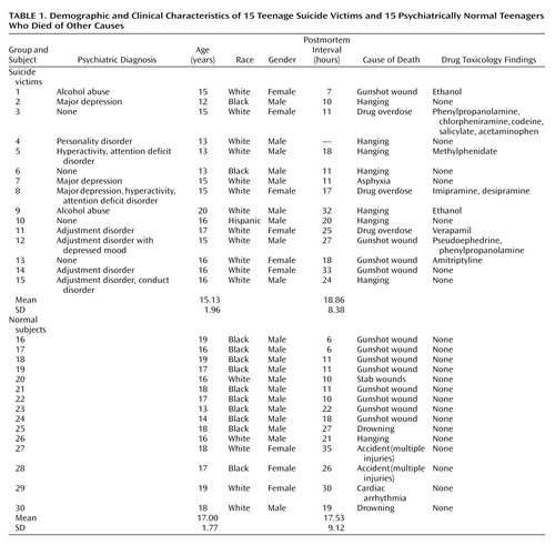
The demographic characteristics, postmortem intervals, and toxicology of the suicide victims and normal subjects are shown in Table 1. There were no significant differences in age or postmortem interval between the suicide victims and the normal subjects. We did not observe any significant effects of age on [125I]LSD binding (r=0.24, df=28, p=0.49), protein expression of 5-HT2A receptor immunolabeling (r=0.26, df=28, p=0.16), or mRNA expression levels (r=0.11, df=28, p=0.56) in the prefrontal cortex. Similarly, there were no significant effects of postmortem interval on [125I]LSD binding (r=0.11, df=28, p=0.55), mRNA expression levels (r=0.17, df=28, p=0.37), or protein expression levels (r=0.19, df=28, p=0.31) in the prefrontal cortex. There were no significant correlations between gender and [125I]LSD binding (r=0.07, df=28, p=0.71), mRNA levels (r=0.03, df=28, p=0.88), or protein expression (r=0.03, df=28, p=0.88) in the prefrontal cortex. As in the prefrontal cortex, we did not find any significant effects of age, postmortem interval, or gender on [125I]LSD binding, mRNA expression, or protein levels of 5-HT2A receptors in the hippocampus or nucleus accumbens. Although we did not observe any significant correlation between age and 5-HT2A receptors, such a relationship could not be ruled out, since the age range of our subjects was so narrow (i.e., 13 to 19 years).
The group of suicide victims was composed of one Hispanic, two blacks, and 12 whites; the group of normal subjects was composed of 10 blacks and five whites. No significant effect of race was observed on any of the measures of 5-HT2A receptors in either the prefrontal cortex, hippocampus, or nucleus accumbens.
[125I]LSD Binding Sites
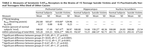
Earlier studies have examined 5-HT2A receptors by means of radioligand binding techniques with different radioligands. Since a higher level of 5-HT2A receptor binding sites has been reported in the brains of adult suicide victims, it was of interest to examine whether similar changes were present in the prefrontal cortex of teenage suicide victims. We determined 5-HT2A binding sites by Scatchard analysis, using [125I]LSD as the ligand and a saturation isotherm and Scatchard plot for a normal subject and a suicide victim, as shown in Figure 1. After analyzing the binding data, we observed that the mean Bmax value of [125I]LSD binding was significantly greater in this brain area of teenage suicide victims than in this area of normal subjects, without any significant difference in KD values (Table 2).
Levels of mRNA Expression
We studied earlier the distribution of 5-HT2A receptor mRNA in various areas of the human brain and observed that 5-HT2A receptor mRNA levels are highly expressed in various cortical areas, the hippocampus, amygdala, and nucleus accumbens (14); therefore, we next quantitated 5-HT2A receptor mRNA in the prefrontal cortex, hippocampus, and nucleus accumbens obtained from teenage suicide victims and normal subjects, using the quantitative reverse transcription polymerase chain reaction technique. A representative autoradiogram of the gel electrophoresis of 5-HT2A receptors in the prefrontal cortex is given in Figure 2A. As expected, we observed the amplification product arising from the mRNA template at 405 bp and the corresponding digestion products arising from the cRNA at 208+197 bp. As shown in Table 2, we found that the mean 5-HT2A mRNA levels were significantly greater in the prefrontal cortex of the 15 teenage suicide victims than in the 15 normal subjects. We also found that the mean mRNA levels in the hippocampus of the suicide victims were significantly higher than in the normal subjects, as shown in Table 2.
To determine if the higher levels of 5-HT2A mRNA observed in the prefrontal cortex and hippocampus were generalized abnormalities or were specific to certain relevant areas of the brain, we determined 5-HT2A receptor mRNA levels in the nucleus accumbens, which also is rich in 5-HT2A receptor mRNA (14). However, the mean 5-HT2A receptor mRNA levels in the nucleus accumbens of suicide victims were not significantly different from those of the normal subjects, as shown in Table 2, indicating that the higher 5-HT2A receptor mRNA in suicide victims is specific to certain brain areas, such as the prefrontal cortex and hippocampus.
Immunolabeling of 5-HT2A Receptors
To further examine if the changes in the gene transcription (mRNA levels) of 5-HT2A receptors also occur at the translational level, we sought to determine the immunolabeling of 5-HT2A receptor protein, using a specific antibody. A representative autoradiogram showing a Western blot of 5-HT2A receptors is depicted in Figure 2B, which shows that the levels of 5-HT2A receptor protein are much higher in the prefrontal cortex from three teenage suicide victims than in the brains of three normal subjects. Comparison of the mean 5-HT2A receptor protein levels in the prefrontal cortex of 15 suicide victims and 15 normal subjects revealed that the protein expression levels of 5-HT2A receptors were significantly higher in these suicide victims than in the normal subjects (Table 2). The mean 5-HT2A protein levels were also significantly higher in the hippocampus, but not in the nucleus accumbens, of the suicide victims compared with the normal subjects (Table 2), suggesting that abnormal 5-HT2A protein levels are specific to the prefrontal cortex and hippocampus and that this larger amount is not a generalized abnormality, since these levels appeared to be normal in the nucleus accumbens of the suicide victims.
Immunocytochemical Localization
In accordance with previously published reports (16–18) making use of immunogold labeling, we found the expression of 5-HT2A receptors to be most dense in pyramidal neurons in layers III, V, and VI and their apical dendrites (Figure 3). Immunogold labeling for 5-HT2A receptors was also observed in a small subgroup of γ-aminobutyric acid (GABA)-ergic neurons, and a moderate expression was also found in glial cells. To localize the changes in the expression density of 5-HT2A receptors at the cellular level, we quantified the number of immunogold particles on pyramidal cell somata and in soma-free regions of the neuropil. Because of its localization in only a subset of GABA-ergic interneurons, the expression density of 5-HT2A receptors was not quantified on this cell type. Figure 3 shows low-magnification photomicrographs of 20-μm sections of Brodmann’s area 9 from a normal teenage subject and a teenage suicide victim that were immunogold-labeled for 5-HT2A receptors. Higher magnification of layer V pyramidal neurons shows a higher expression density of 5-HT2A receptors in the teenage suicide victim than in the normal subject. The mean expression levels of 5-HT2A receptors were significantly higher in the pyramidal cells of layer V of the teenage suicide victims (N=9) than in the normal subjects (N=9), whereas no changes in expression levels of 5-HT2A receptors were found in the pyramidal cells of other cortical layers (layers III and VI) or in the surrounding neuropil (i.e., layers IV, V, and VI) (Figure 4). The mean values of positive neurons of 5-HT2A receptor (number of gold particles/100 μm2) in cortical layers in the normal subjects and the suicide victims were as follows: pyramidal cells (layer III—normal subjects: mean=537.23 gold particles/100 μm2, SD=143.51, suicide victims: mean=502.92, SD=85.91; layer V—normal subjects: mean=341.75, SD=54.06, suicide victims: mean=542.73, SD=82.34; layer VI—normal subjects: mean=442.87, SD=74.75, suicide victims: mean=445.62, SD=78.69); neuropil (layer IV—normal subjects: mean=476.90, SD=107.19, suicide victims: mean=437.85, SD=78.69; layer V—normal subjects: mean=436.62, SD=91.75, suicide victims: mean=381.27, SD=58.70; layer VI—normal subjects: mean=413.82, SD=68.65, suicide victims: mean=396.56, SD=70.80). Thus, it appears that the higher expression of 5-HT2A receptors in the postmortem brains of teenage suicide victims is restricted to the pyramidal cells of the somata of cortical layer V.
Several studies (7) have suggested an association between mental disorders and serotonin receptors in postmortem brains of adult suicide victims. Therefore, we examined the association between 5-HT2A receptors and mental illness in teenage suicide victims. Five suicide victims had no history of mental illness, eight had a history of mental illness (depression, adjustment disorder), and two had a history of alcohol abuse. Although there were no significant differences in any of the 5-HT2A receptor measures, as shown in Figure 5 and Figure 6, in teenage suicide victims with or without mental illness in any of the brain areas studied, the mean mRNA (attomoles/μg of total RNA) in the prefrontal cortex tended to be higher in the suicide victims with mental illness (mean=1197.00 attomol/μg, SD=604.11) than in the suicide victims with no mental illness (mean=883.80 attomol/μg, SD=372.74). Because of the small group size, we could not examine the relationship between specific mental illness, such as depression, and 5-HT2A indices. Our results, however, do indicate that 5-HT2A receptor expression is higher in the prefrontal cortex of suicide victims, independent of a history of mental disorders.
Discussion
We have examined the role of 5-HT2A receptors in teenage suicide, using postmortem brain samples. Although it is not clear if the etiology or the factors underlying teenage suicide are similar to those found in adult suicide, several factors lend credence to a relationship between serotonin and teenage suicide. Most teenage suicide is driven by impulsive or aggressive behavior, stress, or anxiety, which have been shown to be related to abnormalities in serotonergic mechanisms (6); hence, it is quite likely that teenage suicide may be related to abnormalities in serotonergic mechanisms.
We found a striking difference in 5-HT2A receptors between teenage suicide victims and normal subjects. First, we observed that 5-HT2A receptor binding sites are more numerous in the prefrontal cortex of teenage suicide victims than in normal subjects. The observation of a higher level of 5-HT2A receptors in the prefrontal cortex of teenage suicide victims is similar to that observed in adults in previous studies (7). However, we also observed a higher gene expression of 5-HT2A receptors and of its cognate protein in the prefrontal cortex and hippocampus, but not in the nucleus accumbens, in the postmortem brains of teenage suicide victims than in normal subjects. These differences were not related to postmortem interval or age, and the absence of correlation between age and 5-HT2A receptors could be due to the narrow age range of our subjects.
Since it has recently been reported that 5-HT2A receptors are highly expressed in the pyramidal neurons in cortical layers III, V, and VI of the primate and rat brain (18, 19), we determined whether the observed higher protein levels of 5-HT2A receptors were restricted to these specific areas or whether this change was localized throughout all the cortical layers. Similar to findings in the primate brain, in this study we found that 5-HT2A receptors were densely expressed in pyramidal neurons in layers III, V, and VI and to a lesser extent in the surrounding neuropil. Our comparison of the expression of 5-HT2A receptors in various cortical layers of the brains of teenage suicide victims and normal subjects led to an interesting observation that 5-HT2A receptor protein expression is higher only in the pyramidal cells of cortical layer V but not in cortical layers III or VI or in the surrounding neuropil. Our findings thus show that a greater number of active binding sites of 5-HT2A receptors is not merely a consequence of more binding sites but also that gene expression contributes to this greater number. More important, this higher level is restricted to a specific cortical layer. This is an important observation because pyramidal neurons occupy a unique position as they modulate and integrate neuronal functions mediated by serotonergic, glutamatergic, GABA-ergic, and dopaminergic systems (20). It is also important because it has been shown that the soma and dendrites of pyramidal neurons of layer V receive synaptic contact from dopamine terminals and GABA-containing neurons. Since it has been shown that GABA-ergic mechanisms, including benzodiazepine receptors, may be altered in the postmortem brains of suicide victims (21), it is quite possible that higher levels of 5-HT2A receptors in pyramidal cells may cause an imbalance between the 5-HT2A and the GABA-ergic systems and may play an important role in suicidal behavior. Jakab and Goldman-Rakic (20) wrote that “the apical dendritic field proximal to the pyramidal cells soma is the hot spot for 5-HT2A receptor mediated physiological actions relevant to neuronal and psychotic functional states of the cerebral cortex.” They also observed from in vivo recordings in the prefrontal cortex of monkeys that 5-HT facilitates excitatory neurotransmission in the pyramidal neurons engaged in information processing.
Another important observation of this study pertains to the higher 5-HT2A receptor expression specifically in the prefrontal cortex and hippocampus but not in the nucleus accumbens of suicide victims. The prefrontal cortex and hippocampus have been implicated in emotion, stress, and cognitive functions; the nucleus accumbens has been associated with craving, drug dependence, and psychomotor-stimulated behavioral sensitization (22). Both emotion and stress play important roles in suicidal behavior, especially in youth. The higher 5-HT2A receptor indices specifically in the prefrontal cortex and hippocampus thus further strengthen the association of higher levels of 5-HT2A receptors and suicidal behavior.
5-HT2A receptors have been previously studied in the postmortem brains of adult suicide victims with depression and also in nonsuicidal schizophrenia victims. Whereas higher levels of 5-HT2A receptors have been reported in depressed suicide victims (9), a lower number of 5-HT2A receptor binding sites and lower mRNA levels have been reported in patients with schizophrenia (23). From these studies, it is not clear if higher 5-HT2A receptor levels are associated with suicide, depression, or suicidal schizophrenia, since insufficient data exist for suicidal schizophrenia patients. In our study of teenage suicide victims, we examined the relationship of suicide and/or mental disorders with 5-HT2A receptors. Since the numbers of victims with specific psychiatric diagnoses was small, we grouped all victims with a history of mental disorders together, including victims with mood and/or adjustment/conduct disorders. A significant number of victims had no history of mental disorders. Since we did not find any significant differences in any of the 5-HT2A receptor measures (mRNA, protein expression, or binding) between suicide victims with or without a history of mental disorders in any of the brain areas we studied, it appears that the higher level of 5-HT2A receptors is specific to suicide and most likely independent of psychiatric diagnosis. However, these 5-HT2A receptor levels were still significantly higher in both of these groups, compared with the normal subjects. Although this finding is consistent with our previous report that the higher level of platelet 5-HT2A receptors observed in suicidal victims is independent of diagnosis (11), confirmation in a larger number of suicide victims with specific diagnoses of psychiatric disorders is needed. It is also possible that in teenage suicide victims, higher levels of 5-HT2A receptors may be related to impulsive-aggressive behavior. However, this could not be ascertained in this study group, since no data regarding aggressive/impulsive behavior in these victims were available.
The reasons for the higher levels of 5-HT2A receptors in suicide victims are not known, and it is unclear if higher levels of 5-HT2A receptors in the brains of suicide victims indicate a higher vulnerability for suicide. In a recent study, Du et al. (24) examined the 5-HT2A receptor gene in platelets obtained from suicidal and nonsuicidal depressed patients and normal comparison subjects. They found a significant association between the 102C allele in the 5-HT2A receptor gene and major depression, particularly in patients with suicidal ideation. Since the same gene encodes both brain and platelet 5-HT2A receptors (25), our observation of higher 5-HT2A receptor levels in the prefrontal cortex may represent a greater genetic vulnerability for suicide.
However, it is possible that the higher 5-HT2A receptor levels in suicide victims is secondary to changes in other systems, such as abnormalities in the hypothalamic-pituitary-adrenal (HPA) axis. Abnormal HPA axis function and higher levels of cortisol have been consistently observed in patients with depression (26, 27) and suicidal behavior (28, 29). Interaction between the HPA axis and the serotonergic system has also been clearly demonstrated (reviewed by Chauloff [30], Kuroda et al. [31], Fernandes et al. [32], and Mikuni et al. [33]). Moreover, it has been shown that the administration of dexamethasone or corticosterone causes an increase in 5-HT2A receptor levels in the rat cortex (31). Recently, Farisse et al. (34) demonstrated that transgenic mice bearing a transgene coding for glucocorticoid receptor antisense mRNA, which partially blocks glucocorticoid receptor expression, show lower glucocorticoid receptor numbers and a higher level of 5-HT2A receptors. Since cortisol levels are higher in patients with depression and during periods of stress or anxiety, the higher levels of 5-HT2A receptors in the brain of suicide victims may be due to an abnormal HPA axis. It is also quite possible that genetic and environmental factors (higher cortisol level) may in combination cause up-regulation of cortical 5-HT2A receptors.
In summary, this is the first study to our knowledge that demonstrates higher levels of gene expression of 5-HT2A receptors and of the encoded 5-HT2A receptor protein in the prefrontal cortex of suicide victims (either teenage or adult); it shows that these higher levels are restricted to the pyramidal cells of layer V, which are considered a “hotspot” for 5-HT2A receptor-mediated physiological actions. This higher level of 5-HT2A receptors may play a vital role in suicidal behavior and may represent a greater risk for suicide.
 |
 |
Received Nov. 29, 2000; revisions received April 16 and Aug. 1, 2001; accepted Aug. 27, 2001. From the Psychiatric Institute, Department of Psychiatry, University of Illinois at Chicago; and Maryland Psychiatric Research Center, Baltimore, Md. Address reprint requests to Dr. G.N. Pandey, Psychiatric Institute, University of Illinois at Chicago, 1601 W. Taylor St., Chicago, IL 60612; [email protected] (e-mail). Supported by an NIMH grant (MH-48153) to Dr. Pandey. Brain collection was supported in part by an NIMH grant (MH-40279) to Dr. Carpenter. The authors thank Dr. John Smialek, chief medical examiner, and Dr. Dennis Chute, assistant medical examiner, for the collection of brain samples; Boris Lapidus, M.D., and Christopher Grove for dissections; Terri U’Prichard for help with psychological autopsies; and Barbara Brown, Janardhana R. Jonnalagadda, and Miljana Petkovic for technical assistance.

Figure 1. Representative Saturation Isotherms of [125I]LSD Binding to 5-HT2A Receptors in the Brains of a Teenage Suicide Victim and a Psychiatrically Normal Teenager Who Died of Another Causea
aEach point is the mean of triplicate determinations. For this particular experiment, the binding indices for the normal subject were as follows: KD=0.32 nM, Bmax=298 fmol/mg protein, r=0.97. For the suicide victim, they were as follows: KD=0.51 nM, Bmax=425 fmol/mg protein, r=0.99.

Figure 2. Representative Gel Electrophoresis Depicting 5-HT2A Receptor mRNA in the Prefrontal Cortex of a Psychiatrically Normal Teenager Who Died From a Nonsuicide Cause (A)a and Representative Western Blots From the Prefrontal Cortex of Three Teenage Suicide Victims and Three Normal Subjects (B)b
aTotal RNA (1 μg) and decreasing concentrations of cRNA were reverse transcribed and amplified with polymerase chain reaction by using primers specific for the 5-HT2A receptor. The samples were then digested with BgIII restriction enzyme and electrophoresed on 1.5% agarose gel. The upper band depicts 5-HT2A receptor RNA (405 bp). The lower bands depict BgIII-digested internal standard cRNA (208+197).
bProtein samples (30 μg) were resolved with 7.5% acrylamide gel electrophoresis and transferred to nitrocellulose membrane, which was then incubated with 5-HT2A receptor antibody. The membranes were stripped and reprobed with β-actin antibody. The ratio of optical densities of the bands of 5-HT2A receptors to β-actin were calculated.

Figure 3. Low-Magnification Photomicrographs of 20-μm Brain Sections Showing the Six Cortical Layers of the Prefrontal Cortex, Immunogold-Labeled for Serotonin 5-HT2A Receptors, and High-Magnification Photomicrographs of Pyramidal Cells in Brain Layer V From a Teenage Suicide Victim and a Psychiatrically Normal Teenager Who Died of Another Causea
aNote the higher level of 5-HT2A immunogold labeling on the somata, proximal, and apical dendrites of pyramidal cells in layer V of the prefrontal cortex of the suicide victim.

Figure 4. Mean Number of Gold Particles Labeling Serotonin 5-HT2A Receptors in 100-μm2 Sections From Cortical Layers III–VI of Teenage Suicide Victims and Psychiatrically Normal Teenagers Who Died of Other Causes
aSignificant difference between groups (F=21.82, df=1, 14, p<0.0001).

Figure 5. Effects of Mental Disorders on 5-HT2A Receptor Measures in the Prefrontal Cortex of Teenage Suicide Victims
aSignificant difference between groups (F=3.75, df=3, 26, p<0.03).
bSignificant difference between groups (F=13.89, df=3, 26, p<0.001).
cSignificant difference between groups (F=3.45, df=3, 26, p<0.04).

Figure 6. Effects of Mental Disorders on mRNA and Protein Levels of 5-HT2A Receptors in the Hippocampus of Teenage Suicide Victims
aSignificant difference between groups (F=3.86, df=3, 19, p<0.03).
bSignificant difference between groups (F=4.72, df=3, 19, p<0.02).
1. Moscicki EK, O’Carroll P, Rae DS, Locke BZ, Roy A, Regier DA: Suicide attempts in the Epidemiologic Catchment Area study. Yale J Biol Med 1988; 61:259-268Medline, Google Scholar
2. Botsis AF, Soldatos CR, Stefanis CN: Suicide: Biopsychosocial Approaches. Amsterdam, Elsevier, 1997Google Scholar
3. Cross CK, Hirschfeld RMA: Psychosocial factors and suicidal behavior: life events, early loss, and personality. Ann NY Acad Sci 1986; 487:77-89Crossref, Medline, Google Scholar
4. Brent D: Adolescent psychiatric inpatients’ risk of suicide attempt at 6-month follow-up. J Am Acad Child Adolesc Psychiatry 1993; 32:95-105Crossref, Medline, Google Scholar
5. Apter A: Correlation of suicidal and violent behavior in different diagnostic categories in hospitalized adolescent patients. J Am Acad Child Adolesc Psychiatry 1995; 34:912-918Crossref, Medline, Google Scholar
6. Linnoila VM, Virkkunen M: Aggression, suicidality, and serotonin. J Clin Psychiatry 1992; 53:46-51Medline, Google Scholar
7. Gross-Isseroff R, Biegon A, Voet H, Weizman A: The suicide brain: a review of postmortem receptor/transporter binding studies. Neurosci Biobehav 1998; 22:653-661Crossref, Medline, Google Scholar
8. Mann JJ, Stanley M, McBride PA, McEwen BS: Increased serotonin2 and β-adrenergic receptor binding in the frontal cortices of suicide victims. Arch Gen Psychiatry 1986; 43:954-959Crossref, Medline, Google Scholar
9. Arango V, Ernsberger P, Marzuk PM, Chen JS, Tierney H, Stanley M, Reis J, Mann JJ: Autoradiographic demonstration of increased serotonin 5HT2 and β-adrenergic receptor binding sites in the brain of suicide victims. Arch Gen Psychiatry 1990; 47:1038-1046Crossref, Medline, Google Scholar
10. Hrdina PD, Demeter E, Vu TB, Sotonyi P, Palkovits M: 5HT uptake sites and 5HT2 receptors in brain of antidepressant-free suicide victims/depressives: increase in 5HT2 sites in cortex and amygdala. Brain Res 1993; 614:37-44Crossref, Medline, Google Scholar
11. Pandey GN, Pandey SC, Dwivedi Y, Sharma RP, Janicak PG, Davis JM: Platelet serotonin-2A receptors: a potential biological marker for suicidal behavior. Am J Psychiatry 1995; 152:850-855Link, Google Scholar
12. Hrdina PD, Bakish D, Chuzdik J, Ravindran A, Lapierre YD: Serotonergic markers in platelet of patients with major depression: upregulation of 5HT2 receptors. J Psychiatry Neurosci 1995; 20:11-19Medline, Google Scholar
13. Spitzer RL, Williams JBW, Gibbon M, First MB: The Structured Clinical Interview for DSM-III-R (SCID), I: history, rationale, and description. Arch Gen Psychiatry 1992; 49:624-629Crossref, Medline, Google Scholar
14. Salzman S, Endicott J, Clayton P, Winokur G: Diagnostic Evaluation After Death (DEAD). Rockville, Md, National Institute of Mental Health, Neuroscience Research Branch, 1983Google Scholar
15. Dwivedi Y, Pandey GN: Quantitation of 5HT2A receptor mRNA in human postmortem brain using competitive RT-PCR. Neuroreport 1998; 9:3761-3765Crossref, Medline, Google Scholar
16. Hamada S, Senzaki K, Hamaguchi-Hamada K, Tabuchi K, Yamamoto H, Yamamoto T, Yoshikawa S, Okano H, Okado N: Localization of 5HT2A receptor in rat cerebral cortex and olfactory system revealed by immunohistochemistry using two antibodies raised in rabbit and chicken. Mol Brain Res 1998; 54:199-211Crossref, Medline, Google Scholar
17. Jakab RL, Goldman-Rakic PS: Segregation of serotonin 5HT2A and 5HT3 receptors in inhibitory circuits of the primate cerebral cortex. J Comp Neurol 2000; 417:337-348Crossref, Medline, Google Scholar
18. Willins DL, Deutch AY, Roth BL: Serotonin 5HT2A receptors are expressed on pyramidal cells and interneurons in the rat cortex. Synapse 1997; 27:79-82Crossref, Medline, Google Scholar
19. Aghajanian GK, Marek GJ: Serotonin induces excitatory postsynaptic potentials in apical dendrites of neocortical pyramidal cells. Neuropharmacology 1997; 36:589-599Crossref, Medline, Google Scholar
20. Jakab RL, Goldman-Rakic PS: 5-Hydroxytryptamine2A serotonin receptors in the primate cerebral cortex: possible site of action of hallucinogenic and antipsychotic drugs in pyramidal cell apical dendrites. Proc Natl Acad Sci USA 1998; 95:735-740Crossref, Medline, Google Scholar
21. Pandey GN, Conley RR, Pandey SC, Goel S, Roberts RC, Tamminga CA, Chute D, Smialek J: Benzodiazepine receptors in the postmortem brain of suicide victims and schizophrenic victims. Psychiatry Res 1997; 71:137-149Crossref, Medline, Google Scholar
22. Koob GF: Drugs of abuse: anatomy, pharmacology and function of reward pathways. Trends Pharmacol Sci 1992; 13:177-182Crossref, Medline, Google Scholar
23. Hernandez I, Sokolov BP: Abnormalities in 5HT2A receptor mRNA expression in frontal cortex of chronic elderly schizophrenics with varying histories of neuroleptic treatment. J Neurosci Res 2000; 59:218-225Crossref, Medline, Google Scholar
24. Du L, Bakish D, Lapierre YD, Ravindran AV, Hrdina PD: Association of polymorphism of serotonin2A receptor gene with suicidal ideation in major depressive disorder. Am J Med Genet Neuropsychiatry Genet 2000; 96:56-60Crossref, Google Scholar
25. Cook EH Jr, Fletcher KE, Wainwright M, Marks N, Yan SY, Leventhal BL: Primary structure of the human platelet serotonin 5HT2A receptor: identity with frontal cortex serotonin 5HT2A receptor. J Neurochem 1994; 63:465-469Crossref, Medline, Google Scholar
26. Kathol RG, Jaeckel RS, Lopez JF, Meller WH: Pathophysiology of HPA axis abnormalities in patients with major depression: an update. Am J Psychiatry 1989; 146:311-317Link, Google Scholar
27. Holsber F, Lauer CJ, Schreiber W, Krieg JC: Altered hypothalamic-pituitary-adrenocortical regulation in healthy victims at high familial risk for affective disorders. Neuroendocrinology 1995; 62:340-347Crossref, Medline, Google Scholar
28. Arato M, Banki CM, Bissette G, Nemeroff CB: Elevated CSF CRF in suicide victims. Biol Psychiatry 1989; 25:355-359Crossref, Medline, Google Scholar
29. Lopez JF, Palkovits M, Arato M, Mansour A, Akil H, Watson SJ: Localization and quantification of pro-opiomelanocortin mRNA and glucocorticoid receptor mRNA in pituitaries of suicide victims. Neuroendocrinology 1992; 56:491-501Crossref, Medline, Google Scholar
30. Chauloff F: Regulation of 5HT receptors by corticosteroids: where do we stand? Fundam Clin Pharmacol 1995; 9:219-233Crossref, Medline, Google Scholar
31. Kuroda Y, Mikuni M, Nomura N, Takahashi K: Differential effect of subchronic DEX treatment on serotonin-2 and β-adrenergic receptors in the rat cerebral cortex and hippocampus. Neurosci Lett 1993; 155:195-198Crossref, Medline, Google Scholar
32. Fernandes C, McKittrick CR, File SE, McEwen BS: Decreased 5-HT1A and increased 5-HT2A receptor binding after chronic corticosterone associated with a behavioural indication of depression but not anxiety. Psychoneuroendocrinology 1997; 22:477-491Crossref, Medline, Google Scholar
33. Mikuni M, Kusumi I, Kagaya A, Kuroda Y, Mori H, Takahashi K: Increased 5-HT-2 receptor function as measured by serotonin-stimulated phosphoinositide hydrolysis in platelets of depressed patients. Prog Neuropsychopharmacol Biol Psychiatry 1991; 15:49-61Crossref, Medline, Google Scholar
34. Farisse J, Hery F, Barden N, Hery M, Boulenguez P: Central 5-HT1 and 5-HT2 binding sites in transgenic mice with reduced glucocorticoid receptor number. Brain Res 2000; 862:145-153Crossref, Medline, Google Scholar