Human White Matter Atlas
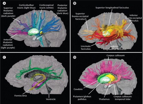
The axonal membrane and the myelin sheets around axons restrict water diffusion perpendicular to the fiber orientation in white matter but not along the axon’s main axis, which leads to anisotropic diffusion of water. Over the last decade, the quantitative measurement of this anisotropy by using what has been called diffusion tensor imaging (DTI) has become well established in research applications, revealing the macroscopic three-dimensional architecture of white matter and the constituent white matter tracts. The depiction of white matter architecture in orientation-based color coding is a visualization approach in which the image brightness depicts diffusion anisotropy and a red/green/blue color scheme indicates tract orientation. Using a multiple region-of-interest (ROI) approach that exploits existing anatomic knowledge of tract trajectories, white matter bundles of interest can be reconstructed non-invasively in vivo . We have created a human white matter brain atlas, consisting of projection, association, and commissural fibers as well as brainstem fibers, examples of which are depicted here.

Figure 1. These figures, taken from a human white matter brain atlas (Mori S, Wakana S, Nagae-Poetscher LM, van Zijl PCM: MRI Atlas of Human White Matter. Amsterdam, Elsevier, 2005), are left lateral views from three-dimensional depictions of fiber pathways, which were developed from normal DTI images. The thalamus, caudate, and putamen and globus pallidus are depicted for anatomic orientation. In box A (Projection and Thalamic Fibers), the anterior, superior, and posterior thalamic radiation derive from the thalamus. The corticobulbar and corticospinal tracts form the projection system. In box B (Association Fibers), the superior and inferior longitudinal faciculus and the superior and inferior fronto-occipital faciculus are organized around the basal ganglia. In box C (Limbic System Fibers), the cingulum, fornix, and stria terminalis fibers are depicted; the ventricular system around which limbic fibers are organized is dark gray. In box D (Callosal Fibers), corticocortical connections pass through the corpus callosum. The subset of these tracts that project to the temporal lobe are pink.


