Neural Networks: Neural Systems II
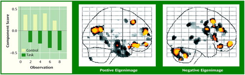
The performance of a cognitive task by a human subject requires an integrated, distributed network of brain regions. However, for a particular task that network may not be known. Eigenimage analysis applied to functional imaging data can suggest data-driven (hypothesis-generating) networks that could subserve the cognitive activity performed during the scanning session. Through a principal-components-type analysis, this technique transforms both the spatial and temporal aspects of the data into independent patterns of functional connectivity. The above image is from an eigenimage analysis of data collected in a [15O]H2O positron emission tomography study that consisted of eight alternating task and control conditions. The task performed during the scan was a visual discrimination of figure height (involving a decision); the control condition provided comparable visual and motor stimuli (involving recognition). This eigenimage has positive loadings (involved in the recognition) primarily in the right and left temporal lobes and negative loadings (involved in the decision) in the cerebellum and right middle frontal lobe. As depicted by the figure, the positive eigenimage is predominant in the control condition while the negative eigenimage is predominant in the discrimination task, with increasing weights associated with later task scans. These data, independently derived, show that activity in a tract from the occipital cortex through to the inferior temporal lobe subserves visual recognition (control condition), while the middle frontal and cerebellar areas subserve decision making (task). The strength of this approach is that it allows for multiple orthogonal (independent) patterns to describe the functional connectivity of the data. This single eigenimage accounts for 51% of the variance-covariance structure of the data. Since the patterns from eigenimage analysis are completely driven by the data, they need to be replicated in a hypothesis-testing experiment. However, this approach can suggest patterns of functional connectivity involving the whole brain over time.
Address reprint requests to Dr. Tamminga, Maryland Psychiatric Research Center, University of Maryland, P.O. Box 21247, Baltimore, MD 21228. Image is courtesy of Dr. Medoff.
Saggital Images
The two saggital images are projections of all significant pixels compressed into two dimensions. The graph depicts the temporal component of the eigenimage analysis, with the regions in the positive image associated with recognition (control condition) and the regions in the negative image associated with decision making (discrimination task).