The Overlapping Neurobiology of Induced and Pathological Anxiety: A Meta-Analysis of Functional Neural Activation
Abstract
Objective:
Although anxiety can be an adaptive response to unpredictable threats, pathological anxiety disorders occur when symptoms adversely affect daily life. Whether or not adaptive and pathological anxiety share mechanisms remains unknown, but if they do, induced (adaptive) anxiety could be used as an intermediate translational model of pathological anxiety to improve drug development pipelines. The authors therefore compared meta-analyses of functional neuroimaging studies of induced and pathological anxiety.
Methods:
A systematic search of the PubMed database was conducted in June 2019 for whole-brain functional MRI articles. Eligible articles contrasted either anxious patients to control subjects or an unpredictable-threat condition to a safe condition in healthy participants. Five anxiety disorders were included: posttraumatic stress disorder, social anxiety disorder, generalized anxiety disorder, panic disorder, and specific phobia. A total of 3,433 records were identified, 181 articles met selection criteria, and the largest subset of task type was emotional (N=138). Seed-based d-mapping software was used for all analyses.
Results:
Induced anxiety (N=693 participants) and pathological anxiety (N=2,554 patients and 2,348 control subjects) both showed increased activation in the left and right insula (coordinates, 44, 14, −14 and −38, 20, −8; k=2,102 and k=1,305, respectively) and cingulate cortex/medial prefrontal cortex (−12, −8, 68; k=2,217). When the analyses were split by disorder, specific phobia appeared the most, and generalized anxiety disorder the least, similar to induced anxiety.
Conclusions:
This meta-analysis indicates a consistent pattern of activation across induced and pathological anxiety, supporting the proposition that some neurobiological mechanisms overlap and that the former may be used as a model for the latter. Induced anxiety might nevertheless be a better model for some anxiety disorders than others.
Anxiety disorders constitute the most prevalent mental health condition (1), with a lifetime prevalence of 17% (2), resulting in significant individual and social impairment (1) and a considerable overall burden of disease, ranking ninth among causes of years lived with disability in the world in 2015 (3). Response rates to existing treatments usually range between 40% and 60% (4), which leaves a large number of people with debilitating symptoms and a high probability of relapse (5).
Development of new treatments for symptoms of anxiety has stagnated for several decades (6), however, partly as a result of the difficulty of establishing robust translational links between models of fear and anxiety in rodents and clinical anxiety in humans. It has recently been argued, therefore, that models of anxiety (as defined by aversive anticipation and apprehension of perceived potential but unpredictable threats) in healthy humans could help us bridge this gap and facilitate therapeutic progress (7).
More precisely, using the same techniques to induce anxiety in healthy individuals and animal models should enable us to both better understand the neurobiological basis of anxiety and provide an intermediate route to screen the efficacy of candidate interventions prior to full clinical trial (8). This experimental approach is possible because anxiety, perhaps uniquely among psychiatric symptoms, is also an adaptive behavior with a benefit to survival. Anxiety enhances vigilance to threat and primes defense mechanisms (9), which allows the individual to react faster in dangerous situations. It occurs naturally in every individual—when walking down a dark alley at night, for instance. This adaptive anxiety can be reliably induced in healthy individuals in the laboratory by exposing them to unpredictable threat of rare electrical shocks. This approach is well validated (10) and is reliable both for self-report and task performance (11), and, critically, it is also fully translational: a close paradigm is used in animal models (12). A growing body of literature shows that, in addition to clear increases in subjective and physiological reports of anxiety (13), threat of shock results in cognitive and psychophysiological changes mirroring pathological anxiety (14–16).
Induced and pathological anxiety therefore overlap at the level of symptoms, as both promote functions and states that promote harm avoidance. What remains insufficiently explored, however, is the extent to which underlying neurobiological mechanisms overlap, and whether ostensibly similar symptoms are driven by dissociable underlying mechanisms. Critically, for the “experimental psychopathology” (7) approach to be valid, the assumption that induced anxiety evokes (at least some of) the same neurobiological mechanisms as pathological anxiety must be met, particularly on emotion-related paradigms, where the literature suggests that they lead to similar changes in cognitive performance (14).
Induced anxiety via unpredictable threat paradigms has been shown to involve brain regions involved in emotional-processing, decision-making, and reward circuitry, such as the amygdala, anterior cingulate cortex, medial prefrontal cortex, bed nucleus of the stria terminalis (BNST), insula, and striatum (17–21), but this has not been systematically meta-analyzed. Meta-analyses and systematic reviews exploring the neural circuitry of different anxiety disorders suggest that many of the same regions have been implicated (22–25); however, differences across disorders have not been investigated in the past decade (26, 27). Meta-analyses of fear conditioning studies, a related experimental model that focuses on predictable (rather than unpredictable) threats, have also reported resulting hyperactivation in the dorsal anterior cingulate cortex and left and right anterior insula (28, 29), and a recent meta-analysis investigated shared neural correlates across mood and anxiety disorders (30). What is lacking, however, is an up-to-date systematic meta-analysis directly assessing the extent to which the neural activation in anxiety disorders in emotion-related paradigms overlaps with that evoked by unpredictable threat–induced anxiety (as opposed to fear conditioning) in the general population—in other words, a systematic assessment of the neurobiological links between induced and pathological anxiety and a quantitative assessment of the “experimental psychopathology” approach to anxiety.
Our aims in this meta-analysis were therefore 1) to investigate the common functional neural activity pattern across induced-anxiety studies, 2) to update disorder-specific maps for five anxiety disorders and examine commonalities across all pathological anxiety brain activity in emotion-related paradigms, and 3) to compare neural patterns of induced anxiety to pathological disorders. We used a coordinate-based meta-analytic whole-brain approach (31) to test the broad prediction that activation patterns overlap across induced and pathological anxiety. This approach has important strengths over conventional activation likelihood meta-analyses, as it uses the effect sizes and enables investigation of voxelwise publication bias (32). Unthresholded group maps were also collected where possible to ensure that the results were as precise as possible. In addition, we explored overlap of our induced-anxiety results with a recent fear-conditioning meta-analysis.
Methods
Literature Search and Article Inclusion
A systematic search was conducted in the PubMed database (all studies published before June 11, 2019, including studies in press) for papers on functional MRI whole-brain blood-oxygen-level-dependent activity reporting contrasts of anxious or depressed patients and control subjects or contrasts of unpredictable-threat and safe conditions. A flowchart of the article selection process is provided in Figure S1 in the online supplement; see the Supplementary Methods section for full details.
A total of 181 publications were identified, comprising 2,911 anxious patients and 2,685 control subjects. To improve consistency across the paradigms used for the contrasts comparing patients with control subjects, articles were then split into broad task categories: emotion (exposure to phobic [e.g., spider images], traumatic [e.g., combat films], socioemotional [e.g., faces], or general strongly aversive stimuli [e.g., loud noises]), attention (sensory detection and focus, go/no-go), decision (strategic planning and calculus, monetary decision making), and memory (working memory encoding and retrieval, learning tasks). The main analyses comparing patients with control subjects were focused on the emotion category (138 articles), which includes 2,554 patients and 2,348 control subjects (of which 27 subjects were depressed [but not anxious] control subjects and the rest healthy) because it was the largest paradigm subset. A total of 693 participants undergoing induction of anxiety were included. Studies of posttraumatic stress disorder (PTSD) using traumatized control subjects (22 articles, 325 patients, 353 control subjects) were not included in the main analysis. Unthresholded maps for 17 of the 138 included articles were obtained (one of the included induced-anxiety studies provided unthresholded maps but did not report coordinates [15 subjects]). See Table S1 in the online supplement for a full description of the samples and included articles.
SDM Meta-Analysis Procedure
Activation and deactivation coordinates, as well as the t-threshold and t-values, were collected from each article for the contrast of interest and entered into the SDM-PSI (32) software package, version 6.12.
Full details are provided in the Supplementary Methods section of the online supplement. Briefly, for each article group, coordinate-based maps were reconstituted and preprocessed with default parameters (20-mm full width at half maximum [FWHM], gray matter mask). This led to the following analyses: 1) a meta-analysis of all induced-anxiety articles, 2) a meta-analysis of all pathological anxiety articles using emotional tasks, 3) convergence analysis of induced anxiety compared with pathological anxiety, 4) separate meta-analyses of the PTSD, social anxiety disorder, generalized anxiety disorder, panic disorder, and specific phobia diagnostic groups of pathological anxiety, and 5) separate convergence analyses of induced anxiety compared with each of the five main diagnostic groups.
Publication bias was assessed for each cluster with Egger’s test implemented in SDM, using, for each cluster, the mean effect size from each study. (See the online supplement for exploratory analyses of the non-emotion tasks in pathological anxiety and of PTSD patients compared with traumatized control subjects.)
An exploratory similarity analysis between our induced-anxiety results and a recent fear-conditioning meta-analysis (28) was also conducted via the NeuroVault comparison tool in the similarity search (chosen map: CS+ vs. CS−, pseudo Z scores). Regional correlations were calculated from a brain-masked 4-mm transformation of the original images.
Results
Findings from 138 papers and 5,595 participants are presented here. See Table S2 in the online supplement for a full list of included papers. All collected coordinates as well as t-value files are available online at https://osf.io/9s32h/. All unthresholded whole-brain activation and convergence maps reported below are available online at https://neurovault.org/collections/6012/.
Pathological Anxiety–Associated Brain Activity
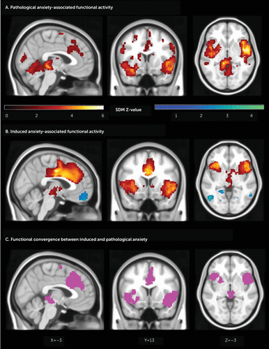
Anxious patients across disorders (N=2,554), compared with control subjects (N=2,348), demonstrated increased activation bilaterally in a cluster encompassing the middle and superior temporal gyri, insula, and inferior frontal gyrus, the left part extending to the amygdala, parahippocampal gyrus, hippocampus, left and right lingual and fusiform gyri, and thalamus (z=5.413 and z=6.156 for left and right clusters, respectively). Increased activation was also found in the anterior and midcingulate and superior medial frontal gyrus (z=3.951). Other clusters of increased activation include the left middle occipital, left postcentral gyrus, left and right caudate, left and right calcarine fissure, left and right precuneus, right supramarginal, left and right superior parietal and superior occipital gyri, right parahippocampal gyrus, left middle frontal gyrus, and supplemental motor area. No clusters of reduced activation were significant. No significant publication bias was revealed by Egger’s test for any peak, including the left (bias=0.34, p=0.519) and right (bias=0.46, p=0.355) superior temporal gyrus/insula/inferior frontal gyrus clusters and the cingulate/medial frontal clusters (bias=0.25 [p=0.666], bias=0.14 [p=0.780], and bias=0.05 [p=0.927], respectively) (Figure 1A and Table 1). Upon specific examination, bilateral increased activation was also found in the periaqueductal gray.

FIGURE 1. Functional activation and convergence for induced and pathological anxiety in a meta-analysis of functional neural activationa
a Panel A shows brain regions differing significantly between threat and safe conditions in induced-anxiety studies (693 participants). Panel B shows brain regions differing significantly between 2,554 anxious patients and 2,348 control subjects across pathological anxiety studies. The SDM Z-value of activation is shown in a red-yellow gradient, and deactivation in a blue-green gradient. Panel C shows convergence of brain regions between induced and pathological anxiety. Converging activation is shown in purple.
| MNI Coordinates (x, y, z) | Voxels | Z | Description | Egger’s Intercept | Egger’s p |
|---|---|---|---|---|---|
| Pathological anxiety | |||||
| –36, 6, –14 | 12,836 | 5.413 | L. insula, inferior frontal gyrus (all), putamen, pallidum, Rolandic operculum, precentral gyrus, postcentral gyrus, middle frontal gyrus, Heschl’s gyrus, superior temporal gyrus pole, superior temporal gyrus, middle temporal gyrus pole, middle temporal gyrus, amygdala, hippocampus, L. and R. parahippocampal gyrus, L. and R. vermis 3–7, L. and R. cerebellum 3–6, cerebellum 8, L. and R. lingual gyrus, L. and R. fusiform gyrus, L. and R. thalamus | 0.34 | 0.519 |
| 48, 4, –14 | 5,701 | 6.159 | R. insula, inferior frontal gyrus (all), superior temporal gyrus pole, superior temporal gyrus, middle temporal gyrus pole, middle temporal gyrus, Heschl’s gyrus, Rolandic operculum, inferior temporal gyrus | 0.46 | 0.355 |
| –10, –2, 68 | 1,203 | 3.951 | L. and R. midcingulate cortex, supplementary motor area, medial frontal gyrus, anterior cingulate cortex, paracentral lobule | 0.25 | 0.666 |
| –36, –74, 30 | 565 | 3.589 | L. middle occipital gyrus, inferior parietal gyrus, superior occipital gyrus, superior parietal gyrus | –0.17 | 0.722 |
| 4, –90, 12 | 333 | 3.362 | Center of calcarine, L. and R. cuneus | 0.39 | 0.397 |
| –26, 22, 40 | 280 | 3.149 | L. middle frontal gyrus, superior frontal gyrus | 0.22 | 0.698 |
| –16, 12, 14 | 202 | 2.975 | L. caudate, thalamus | 0.09 | 0.863 |
| 18, –56, 50 | 180 | 2.667 | R. superior parietal gyrus, precuneus, inferior parietal gyrus | 0.09 | 0.859 |
| 20, –76, 34 | 130 | 2.992 | R. superior occipital gyrus, cuneus | 0.11 | 0.828 |
| 22, –16, –26 | 112 | 3.146 | R. parahippocampal gyrus, hippocampus | 0.20 | 0.685 |
| –14, –58, 58 | 64 | 3.034 | L. precuneus, superior parietal gyrus | 0.26 | 0.611 |
| 8, –66, 18 | 65 | 2.574 | R. calcarine, cuneus | 0.01 | 0.992 |
| 14, 10, 18 | 41 | 2.536 | R. caudate | 0.17 | 0.737 |
| –44, –20, 56 | 37 | 2.557 | L. postcentral gyrus | 0.14 | 0.790 |
| 34, 12, 44 | 30 | 2.279 | R. middle frontal gyrus | 0.03 | 0.948 |
| 58, –32, 40 | 22 | 2.308 | R. supramarginal gyrus | 0.08 | 0.875 |
| 10, –2, 66 | 21 | 2.433 | R. supplementary motor area | 0.05 | 0.916 |
| 12, –28, 40 | 14 | 2.201 | R. midcingulate cortex | 0.14 | 0.780 |
| –8, 6, 38 | 10 | 2.114 | L. midcingulate cortex | 0.05 | 0.927 |
| Induced anxiety | |||||
| 4, 38, 38 | 6,538 | 6.415 | Anterior cingulate cortex, midcingulate cortex, superior medial frontal gyrus | 1.48 | 0.110 |
| 50, 22, 2 | 4,537 | 5.183 | R. insula, inferior frontal gyrus (all), Rolandic operculum, superior temporal gyrus pole | 1.91 | 0.148 |
| –30, 18, –14 | 1,811 | 5.067 | L. insula, inferior frontal gyrus (all), putamen, Rolandic operculum | 1.30 | 0.199 |
| 60, –46, 36 | 1,373 | 4.458 | R. supramarginal gyrus, superior temporal gyrus, angular gyrus | 0.78 | 0.553 |
| 46, 2, 48 | 336 | 3.590 | R. precentral gyrus, middle frontal gyrus | 1.53 | 0.161 |
| –56, –44, 28 | 303 | 3.354 | L. supramarginal gyrus | 1.28 | 0.199 |
| 35, 52, 18 | 153 | 2.500 | R. middle frontal gyrus, superior frontal gyrus | 2.65 | 0.222 |
| –10, –34, –48 | 36 | 3.019 | Possible cerebellum 9, 10 | 0.30 | 0.778 |
| –48, –62, –6 | 1,251 | –4.117 | L. inferior temporal gyrus, middle temporal gyrus, inferior occipital gyrus | –0.32 | 0.752 |
| –56, –22, 46 | 636 | –4.492 | L. postcentral gyrus, inferior parietal gyrus | –0.38 | 0.726 |
| 54, –60, –10 | 559 | –4.102 | R. inferior temporal gyrus, inferior occipital gyrus, middle temporal gyrus, middle occipital gyrus | –0.14 | 0.891 |
| –8, 52, –22 | 478 | –3.267 | L. orbital medial frontal gyrus | –0.11 | 0.932 |
| 42, –44, –16 | 188 | –3.391 | R. fusiform gyrus | –0.25 | 0.826 |
| –12, –64, 12 | 185 | –3.132 | L. calcarine, lingual gyrus | –0.12 | 0.912 |
| –20, –46, 0 | 121 | –3.063 | L. lingual gyrus, fusiform gyrus | –0.32 | 0.769 |
| 26, –62, –8, | 47 | –2.525 | R. fusiform gyrus, lingual gyrus | –0.34 | 0.744 |
| –20, –70, –8 | 44 | –2.697 | L. fusiform gyrus, lingual gyrus | –0.38 | 0.728 |
| –32, –26, –20 | 42 | –2.675 | L. fusiform gyrus, parahippocampal gyrus | –0.42 | 0.695 |
| 12, –70, 16 | 41 | –2.691 | R. calcarine | –0.09 | 0.940 |
| –22, –16, –22 | 37 | –3.052 | L. parahippocampal gyrus, hippocampus | –0.62 | 0.608 |
| 28, –24, –24 | 13 | –2.448 | R. parahippocampal gyrus, fusiform gyrus | 0.08 | 0.945 |
| 18, –78, 14 | 12 | –2.119 | R. calcarine | –0.21 | 0.842 |
| Convergence | |||||
| –12, –8, 68 | 2,217 | Supplementary motor area, midcingulate cortex, medial frontal gyrus (superior), anterior cingulate cortex | |||
| 44, 14, –14 | 2,102 | R. insula, inferior frontal gyrus (all), superior temporal gyrus pole, Rolandic operculum, superior temporal gyrus, putamen | |||
| –38, 20, –8 | 1,305 | L. insula, inferior frontal gyrus (all), putamen, Rolandic operculum, superior temporal gyrus pole | |||
| –4, –22, –10 | 615 | R. thalamus, vermis 3 | |||
| 50, 2, 44 | 183 | R. precentral gyrus, middle frontal gyrus | |||
| 12, –26, 40 | 43 | R. midcingulate cortex | |||
TABLE 1. Whole-brain meta-analysis of induced-anxiety articles in threat compared with safe conditions, and of pathological anxiety articles across disorders, in a meta-analysis of functional neural activationa
Diagnostic Group Analyses
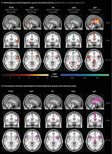
When analyses were broken down into diagnostic groups (Figure 2A), specific phobia (414 patients) showed the three increased activation clusters in the cingulate and left and right inferior frontal gyrus/insula. Panic disorder (263 patients) and PTSD (436 patients) also showed more activation in the left and right insula/superior temporal gyrus but no activation or deactivation in the mid and anterior cingulate cortex. Social anxiety disorder (805 patients) showed activation in the right insula/inferior frontal gyrus/superior temporal gyrus and left amygdala but no activation or deactivation in the cingulate as well. In contrast to the other disorders, generalized anxiety disorder (233 patients) showed deactivation in the cingulate cortex and in the left and right insula. (See Table S3 in the online supplement for full disorder-specific peak information.) No significant publication bias was revealed by Egger’s test for any peak. Increased activation in the periaqueductal gray was found bilaterally in specific phobia and in the left hemisphere in panic disorder.

FIGURE 2. Functional activation and convergence with induced anxiety for each anxiety disorder in a meta-analysis of functional neural activationa
a Panel A shows brain regions differing significantly between anxious patients and control subjects for each anxiety disorder. C=control subjects; GAD=generalized anxiety disorder; P=patients; PD=panic disorder; PTSD=posttraumatic stress disorder; SAD=social anxiety disorder; SpP=specific phobia. The SDM Z-value of activation is shown in a red-yellow gradient, and deactivation in a blue-green gradient. Panel B shows convergence of brain regions between induced anxiety and each anxiety disorder. Converging activation is shown in purple.
Induced Anxiety–Associated Brain Activity
Across participants (N=693), induced anxiety in threat compared with safe conditions demonstrated greater activation in the cingulate and medial frontal cortices (z=6.415), and bilaterally in the inferior frontal gyrus/anterior insula/Rolandic operculum (z=5.183 and z=5.067 for the right and left clusters, respectively). Other areas of increased activation include the left and right supramarginal, right superior temporal, right middle frontal, and right precentral gyri. Reduced activation was found in the left and right parahippocampal gyrus, fusiform, and lingual gyri, as well as in the left and right calcarine fissure, left and right inferior temporal, middle temporal, inferior occipital, left postcentral, and left orbital medial frontal gyri. Egger’s test for publication bias was not significant for any clusters, including the cingulate/medial frontal (bias=1.5, p=0.11) and the left (bias=1.3, p=0.20) and right (bias=1.91, p=0.15) inferior frontal gyrus/anterior insula clusters (see Figure 1B and Table 1). Upon specific examination, the increased activation in the BNST and periaqueductal gray was also found. Restricting the analysis to induction of anxiety via threat of shock did not affect the primary outcome (see Table S4 in the online supplement).
Comparison Between Induced and Pathological Anxiety
When comparing all pathological anxiety with induced anxiety (Figure 1C), we see convergence for increased activation in the left and right insula/inferior frontal gyrus and in the anterior cingulate cortex/midcingulate cortex/superior medial frontal cortex. These clusters were also present, both for activity and convergence, in the complementary 10-mm FWHM analysis. Convergence was also found for bilateral periaqueductal gray activation. Excluding articles reporting any medicated patients or articles using a youth patient sample did not affect the primary outcomes (see Table S5 in the online supplement).
Diagnostic group analyses.
When compared with induced anxiety (Figure 2B), specific phobia showed convergence for increased activation in the cingulate/medial prefrontal and the left and right insula/inferior frontal gyrus/putamen/superior temporal gyrus pole. Panic disorder showed convergence for bilateral insula/inferior frontal gyrus hyperactivation, whereas PTSD was only convergent with induced anxiety for increased activation in the insula/inferior frontal gyrus opercular part, but not for the inferior frontal gyrus triangular or orbital parts. Social anxiety disorder converged in the right insula/inferior frontal gyrus orbital and triangular parts. Generalized anxiety disorder showed very limited overlap with induced anxiety. (See Table S6 in the online supplement for full pairwise convergence peaks.) All clusters mentioned above were also present in the 10-mm FWHM analysis for convergence with induced anxiety, although the left insula contribution to activity in panic disorder was absent. Specific phobia also converged with induced anxiety for bilateral BNST and periaqueductal gray activation.
Overlap of Induced Anxiety With Fear Conditioning
Induced anxiety showed a whole-brain Pearson correlation coefficient (r) of 0.66 with a fear-conditioning meta-analysis. Regional correlations were 0.76 for the putamen, 0.75 for the insula, 0.73 for the frontal lobe, 0.65 for the parietal lobe, 0.57 for the caudate, and 0.54 for the thalamus.
Discussion
Consistent with the hypothesis that induced anxiety may be an experimental psychopathological model of anxiety disorders, induced and pathological anxiety show overlapping neurobiological activations. Specifically, induced anxiety and pathological anxiety both converged for increased activation in the cingulate cortex/medial prefrontal cortex, left and right insula/inferior frontal gyrus, and periaqueductal gray. However, there were also some important dissociations, especially when pathological anxiety was broken down into component disorders, perhaps suggesting that induced anxiety overall might be a model closest to specific phobia and furthest from generalized anxiety disorder.
Induced Anxiety as a Model for Pathological Anxiety
The first thing to note is that induced anxiety evokes activation in the anterior cingulate cortex, midcingulate cortex, medial prefrontal cortex, and insula, as well as activation in the BNST and periaqueductal gray. The insula and cingulate regions have been argued to form part of a “fear-conditioning” circuitry (28) and/or a “salience” network (33) that drives interoception in particular (34). In fact, NeuroVault similarity analysis reveals that our induced-anxiety map shows reasonably high (r≈0.7) correlation with a recent meta-analysis investigating Pavlovian fear conditioning neural correlates (28). The overlapping regions perhaps therefore reflect a shared circuitry that responds to the threats common to anxiety and fear conditioning, with the nonoverlapping circuits perhaps being specific to the spatial/temporal predictability of these threats. Midcingulate cortex electroencephalographic activity has also been reported to play a key role in adapting behavior to uncertainty and to be modulated by anxiety (35). It is therefore possible that these regions contribute to circuitry that (in the case of the cingulate) detects salient environmental stimuli and then promotes behavioral avoidance of threats (via connections to the motor cortex), or (in the case of the insula) detects salient internal change that requires some kind of homeostatic response (e.g., heart rate increase). The overall effect is to reduce the negative impact of potential harms (perhaps in concert as part of a putative “salience” network).
Critically, the same insula and cingulate activations are seen across pooled anxiety disorders in our data, as well as in older meta-analyses (26, 36–38). They may therefore play the same role in pathological anxiety disorders—promoting avoidance responses to salient negative stimuli. Indeed, insula and midcingulate response is thought to be a promising predictor of psychotherapy response (39), which suggests that this circuitry is also important for clinical response (which is largely defined as a reduced avoidance/response to threats).
Thus, induced anxiety holds promise as an intermediate translational model of anxiety disorders (7). In other words, promising new candidate medications might be shown first to modify the effects of threat of shock in animal models (i.e., subjecting animals to unpredictable shocks) (12), and then the effects of threat of shock in healthy humans, before being rolled out in a full-scale clinical trial in anxiety disorders. This would provide greater confidence that the candidate medication targets relevant symptoms and mechanisms and thereby improve the (currently very poor) hit rate of psychiatric drug development (4, 5). This is important because it has been suggested that fear and anxiety in humans can and should both be conceptually segregated across two systems with separate but interacting circuitry: the behavioral and physiological response on the one hand, and the conscious feeling and state on the other hand (40). Conceptually, induction of anxiety via unpredictable threat spans both systems in humans: a conscious but diffuse feeling of anxiety as well as avoidance and physiological defensive arousal. The overlapping activity we observe may therefore be involved in both of these facets, but further work, ideally with identical cognitive tasks across both induced and pathological anxiety, is needed to truly disentangle these important distinctions. At the same time, if very similar manipulations can be used in animal models, this would circumvent, to a certain extent, the problem that it is not possible to measure subjective feeling/states in animal models. In other words, consistent translational manipulations provide a more direct bridge from animal models to human clinical work as well as a means of eventually reconciling disparate anxiety-related systems (40).
Specificity Across Disorders
Although similarities were found with induced anxiety when all the pathological anxiety studies were pooled together, some differences became apparent when studies were split by disorder. Because these results may be confounded by biases in sample sizes and/or cognitive tasks (see the Limitations section below), we must refrain from excessive interpretation, but specific phobia was revealed to be most similar to induced anxiety, showing significant increased activation convergence in the cingulate cortex/medial prefrontal cortex and left and right insula/inferior frontal gyrus as well as in the BNST and periaqueductal gray. PTSD and panic disorder converged with induced anxiety for increased activation in the left and right insula but not for cingulate hyperactivation. Finally, both social anxiety disorder and generalized anxiety disorder had a more complex pattern, the former only converging in the right insula and the latter failing to show convergence for bilateral insula and cingulate hyperactivation. Thus, it may also be that induced anxiety is a better model for some subtypes of pathological anxiety than others.
Overall, the disorder-specific findings may indicate that pathological anxiety mechanisms are diverse and that we should not always assume similarities across disorders. Indeed, with the detection power allowed by the current literature, induction of anxiety, mainly by threat of unpredictable shock, appears to be a very good model for specific phobia, a relatively good model for panic disorder, PTSD, and possibly social anxiety disorder, and less relevant for generalized anxiety disorder at the functional activation level. However, direct comparison with the exact same tasks and the same power in all groups is needed to be confident in this prediction.
Limitations
To our best knowledge, this is the first meta-analysis to investigate functional activity in anxiety induced via unpredictable threat paradigms and compare it with up-to-date meta-analyzed functional activity of pathological anxiety disorders. However, it is important to recognize several limitations.
First, in order to collect sufficient samples in each group of eligible articles, we did not exclude any articles on the basis of male-to-female ratio, age, potential medication, individual comorbidities, and clinical severity—all of which could potentially confound the functional correlates of anxiety in patients or in healthy control subjects.
Second, splitting analyses by anxiety type and disorders (and restricting them to emotional tasks) resulted in varied group sizes, leading to differences in power between diagnosis-specific meta-analyses as well as an overabundance of some task types in some disorders (e.g., symptom provocation in specific phobia). Similarly, at a more global level, the tasks within the induced-anxiety sample are more consistent than the diverse emotion tasks in the pathological anxiety sample. Ultimately, this likely makes all interpretation of the differences between groups less solid than the observed common/shared effects.
Third, although we did restrict analyses to broad task categories, we did not filter our systematic analysis by precise task, because we wanted to examine as many aspects of anxiety as the body of literature allowed. As a result, the tasks used in eligible articles are somewhat diverse. For example, the criteria of our broad emotion task category were exposure to threatening or strongly aversive stimuli (electrical shocks, loud noises, etc.), phobic stimuli (images of spiders, sounds of dental care, etc.), traumatic stimuli (combat-related movies, etc.), or socioemotional stimuli (faces or words with or without emotion, etc.). Since all induced-anxiety articles we selected included strongly aversive threat, they all qualified by definition for this category. Coincidentally, all the eligible specific phobia studies used phobic stimuli in their specific tasks and, as a result, also qualified for the emotion category. Hence, one explanation for the consistency between specific phobia and induced anxiety may be that the symptom provocation tasks used were most similar across these studies. In a broader sense, our inclusive approach where we pool articles across different diagnoses and paradigms will inevitably lead to biases that limit inference. In an ideal world, we would restrict analyses to single tasks, but this would severely limit our detection power. As it stands, our inference is perhaps stronger for the conjunction analyses (where we are seeing similarities in spite of confounders) rather than our difference analyses (where discrepancies may simply be driven by confounders).
Fourth, it is worth noting that we restricted our meta-analysis to articles reporting a whole brain analysis. Unfortunately, for a small number of articles, it was unclear whether the analysis was carried out with homogeneous thresholding across the whole brain or whether some regions of interest were singled out (we would recommend increased clarity in reporting of future research). Notably, some key structures, the amygdala in particular, as well as the BNST and periaqueductal gray, often do not emerge in whole-brain analyses, most of which have a comparatively large cluster threshold. Thus, for the most part, our results did not reflect amygdala activation or deactivation, which are often reported in region-of-interest analyses only.
In summary, this meta-analysis demonstrates that induced anxiety evokes activation of cingulate and insular regions in common with pathological anxiety, which (at least partially) validates the former as an intermediate translational model of the latter. Nevertheless, our findings also indicate functional differences between anxiety disorders, suggesting that induced anxiety might be a better model for some disorders than others.
1 : Prevalence, severity, and comorbidity of 12-month DSM-IV disorders in the National Comorbidity Survey Replication. Arch Gen Psychiatry 2005; 62:617–627Crossref, Medline, Google Scholar
2 : Prevalence and incidence studies of anxiety disorders: a systematic review of the literature. Can J Psychiatry 2006; 51:100–113Crossref, Medline, Google Scholar
3 : Global, regional, and national incidence, prevalence, and years lived with disability for 310 diseases and injuries, 1990–2015: a systematic analysis for the Global Burden of Disease Study 2015. Lancet 2016; 388:1545–1602Crossref, Medline, Google Scholar
4 : Response rates for CBT for anxiety disorders: need for standardized criteria. Clin Psychol Rev 2015; 42:72–82Crossref, Medline, Google Scholar
5 : [Treatment-resistant anxiety disorders: social phobia, generalized anxiety disorder and panic disorder]. Br J Psychiatry 2007; 29(suppl 2):S55–S60 (Portuguese)Crossref, Google Scholar
6 : 50 years of hurdles and hope in anxiolytic drug discovery. Nat Rev Drug Discov 2013; 12:667–687Crossref, Medline, Google Scholar
7 : Modeling anxiety in healthy humans: a key intermediate bridge between basic and clinical sciences. Neuropsychopharmacology 2019; 44:1999–2010Crossref, Medline, Google Scholar
8 : The central extended amygdala in fear and anxiety: closing the gap between mechanistic and neuroimaging research. Neurosci Lett 2019; 693:58–67Crossref, Medline, Google Scholar
9 : Anxiety: an evolutionary approach. Can J Psychiatry 2011; 56:707–715Crossref, Medline, Google Scholar
10 : Assessing fear and anxiety in humans using the threat of predictable and unpredictable aversive events (the NPU-threat test). Nat Protoc 2012; 7:527–532Crossref, Medline, Google Scholar
11 : Towards an emotional “stress test”: a reliable, non-subjective cognitive measure of anxious responding. Sci Rep 2017; 7:40094Crossref, Medline, Google Scholar
12 : Phasic vs sustained fear in rats and humans: role of the extended amygdala in fear vs anxiety. Neuropsychopharmacology 2010; 35:105–135Crossref, Medline, Google Scholar
13 : Models and mechanisms of anxiety: evidence from startle studies. Psychopharmacology (Berl) 2008; 199:421–437Crossref, Medline, Google Scholar
14 : The impact of anxiety upon cognition: perspectives from human threat of shock studies. Front Hum Neurosci 2013; 7:203Crossref, Medline, Google Scholar
15 : Modeling avoidance in mood and anxiety disorders using reinforcement learning. Biol Psychiatry 2017; 82:532–539Crossref, Medline, Google Scholar
16 : Threat of shock and aversive inhibition: induced anxiety modulates Pavlovian-instrumental interactions. J Exp Psychol Gen 2017; 146:1694–1704Crossref, Medline, Google Scholar
17 : Anxiety-potentiated amygdala-medial frontal coupling and attentional control. Transl Psychiatry 2016; 6:
18 : Experiential, autonomic, and neural responses during threat anticipation vary as a function of threat intensity and neuroticism. Neuroimage 2011; 55:401–410Crossref, Medline, Google Scholar
19 : Phasic and sustained fear in humans elicits distinct patterns of brain activity. Neuroimage 2011; 55:389–400Crossref, Medline, Google Scholar
20 : Stress increases aversive prediction error signal in the ventral striatum. Proc Natl Acad Sci USA 2013; 110:4129–4133Crossref, Medline, Google Scholar
21 : How human amygdala and bed nucleus of the stria terminalis may drive distinct defensive responses. J Neurosci 2017; 37:9645–9656Crossref, Medline, Google Scholar
22 : Functional neuroanatomy in panic disorder: status quo of the research. World J Psychiatry 2017; 7:12–33Crossref, Medline, Google Scholar
23 : Neuroimaging in social anxiety disorder: a meta-analytic review resulting in a new neurofunctional model. Neurosci Biobehav Rev 2014; 47:260–280Crossref, Medline, Google Scholar
24 : A review of neuroimaging studies in generalized anxiety disorder: “So where do we stand?” J Neural Transm (Vienna) 2019; 126:1203–1216Crossref, Medline, Google Scholar
25 : Meta-analysis of functional brain imaging in specific phobia. Psychiatry Clin Neurosci 2013; 67:311–322Crossref, Medline, Google Scholar
26 : Functional neuroimaging of anxiety: a meta-analysis of emotional processing in PTSD, social anxiety disorder, and specific phobia. Am J Psychiatry 2007; 164:1476–1488Link, Google Scholar
27 : The neurocircuitry of fear, stress, and anxiety disorders. Neuropsychopharmacology 2010; 35:169–191Crossref, Medline, Google Scholar
28 : Neural signatures of human fear conditioning: an updated and extended meta-analysis of fMRI studies. Mol Psychiatry 2016; 21:500–508Crossref, Medline, Google Scholar
29 : A meta-analysis of instructed fear studies: implications for conscious appraisal of threat. Neuroimage 2010; 49:1760–1768Crossref, Medline, Google Scholar
30 : Shared neural phenotypes for mood and anxiety disorders: a meta-analysis of 226 task-related functional imaging studies. JAMA Psychiatry 2019; 77:1–8Google Scholar
31 : Voxel-wise meta-analysis of grey matter changes in obsessive-compulsive disorder. Br J Psychiatry 2009; 195:393–402Crossref, Medline, Google Scholar
32 : Voxel-based meta-analysis via permutation of subject images (PSI): theory and implementation for SDM. Neuroimage 2019; 186:174–184Crossref, Medline, Google Scholar
33 : The organization of the human cerebral cortex estimated by intrinsic functional connectivity. J Neurophysiol 2011; 106:1125–1165Crossref, Medline, Google Scholar
34 : An insular view of anxiety. Biol Psychiatry 2006; 60:383–387Crossref, Medline, Google Scholar
35 : Frontal midline theta reflects anxiety and cognitive control: meta-analytic evidence. J Physiol Paris 2015; 109:3–15Crossref, Medline, Google Scholar
36 : Decreased intra- and inter-salience network functional connectivity is related to trait anxiety in adolescents. Front Behav Neurosci 2016; 9:350Crossref, Medline, Google Scholar
37 : Uncertainty and anticipation in anxiety: an integrated neurobiological and psychological perspective. Nat Rev Neurosci 2013; 14:488–501Crossref, Medline, Google Scholar
38 : Identification of common neural circuit disruptions in cognitive control across psychiatric disorders. Am J Psychiatry 2017; 174:676–685Link, Google Scholar
39 : Meta-analyses of the neural mechanisms and predictors of response to psychotherapy in depression and anxiety. Neurosci Biobehav Rev 2018; 95:61–72Crossref, Medline, Google Scholar
40 : Using neuroscience to help understand fear and anxiety: a two-system framework. Am J Psychiatry 2016; 173:1083–1093Link, Google Scholar


