Targeting PTSD
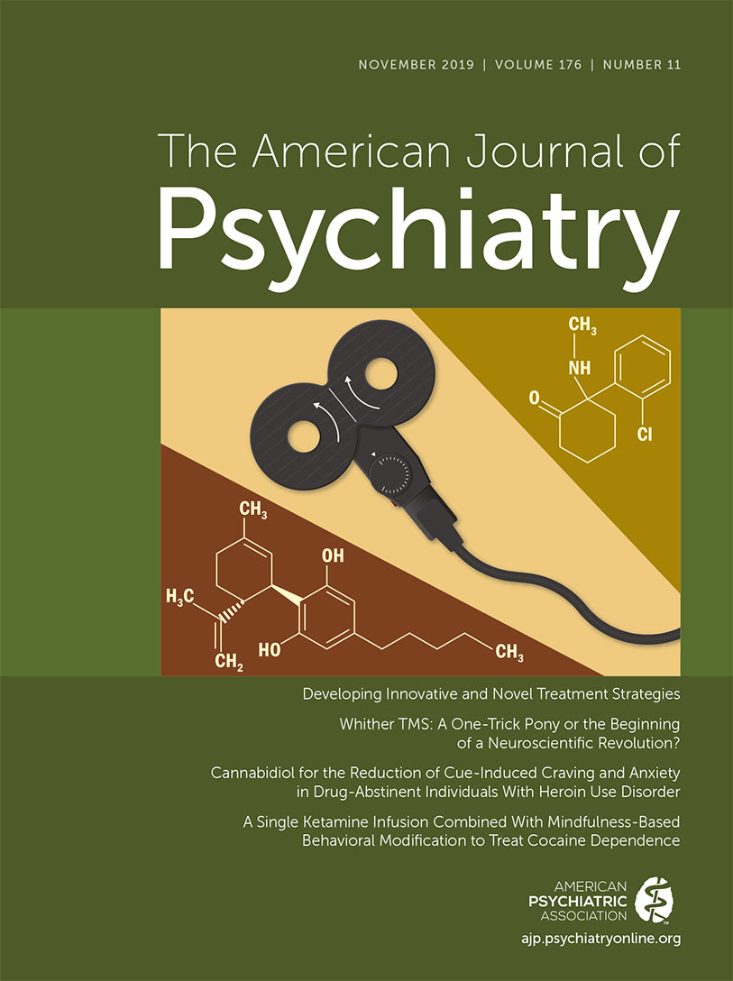
In this issue, Philip et al. (1) detail the results of the first sham-controlled randomized trial using intermittent theta-burst transcranial magnetic stimulation (iTBS) in the treatment of posttraumatic stress disorder (PTSD). The study results support this innovative approach for the treatment of PTSD and demonstrate both the safety and efficacy of iTBS and the use of neuroimaging biomarkers to develop therapeutic treatment targets.
The lifetime prevalence of PTSD is approximately 8% (2). Less than half of patients who meet criteria for PTSD receive appropriate treatment or follow through with evidence-based therapy (3). Psychotherapy, specifically trauma-focused psychotherapies such as prolonged exposure therapy, is very effective. However, these therapies require considerable expertise, time, and resources; almost one-quarter of patients do not complete the treatment course, and up to one-half are left with significant residual symptoms (4). Pharmacotherapy is effective in less than 60% of patients (5), and fewer than one in five patients go into remission (6). A recent randomized controlled trial showed no difference in PTSD treatment outcome between exposure therapy, sertraline, and the combination of exposure therapy and sertraline (7). There is clearly a need for alternative therapies for PTSD (8, 9).
Repetitive TMS (rTMS) was first approved by the U.S. Food and Drug Administration (FDA) for treatment-resistant depression in 2008, and then for treatment-resistant obsessive-compulsive disorder in 2018. Studies of rTMS in PTSD have applied 1–20 Hz rTMS to the right or left dorsolateral prefrontal cortex (DLPFC) or both (reviewed in reference 10). A meta-analysis (10) showed that the overall effect size on PTSD symptoms was large and that the most effective treatment was high-frequency stimulation over the right DLPFC.
iTBS is a form of high-frequency rTMS that delivers brief trains of high-frequency pulses (50 Hz) that are repeated in 200-ms intervals (or 5 Hz, which is in the EEG theta range [4–7 Hz]). Recent iTBS research has focused on treatment-resistant depression. iTBS can deliver effective treatments in 3 minutes, compared with the 37-minute FDA-approved depression protocol for rTMS, with an obvious advantage for both patients and treating physicians. In August 2018, the FDA cleared a 3-minute iTBS protocol in the treatment of depression over the left DLPFC based on the results of the THREE-D (theta-burst versus high-frequency rTMS in patients with depression) inferiority trial, which showed that the iTBS protocol was not inferior to the approved FDA rTMS protocol (11).
iTBS may be a particularly effective form of stimulation in the treatment of PTSD. The theta range is the burst discharge recorded from the hippocampus (12–14), and preclinical studies in mice have shown that TBS can induce long-term potentiation in the lateral amygdala (15). Neurophysiological changes in the amygdala and hippocampus are thought to be integral to the development of the PTSD syndrome.
However, a primary difficulty in treating PTSD and other psychiatric disorders with iTBS (or rTMS) has been the fact that many of the neuroanatomic targets are subcortical (e.g., the amygdala, the subgenual anterior cingulate cortex [sgACC], the hippocampus) and cannot be directly stimulated with these devices. Intermittent theta-burst devices only stimulate cortical tissue that is within a few centimeters under the device. Also, the procedure for determining the location of stimulation relies on establishing a site on the scalp over a target in the parietal lobe, which, when stimulated, causes activation of the abductor pollicis brevis. The site of stimulation is determined by this reference point (e.g., moving the stimulator 6 cm anterior to an area of the DLPFC). Previous research in treatment-resistant depression has shown that this technique frequently misses the DLPFC, which may be associated with a failure to respond to rTMS (16).
A more precise method to determine the effective site of stimulation is to use resting-state functional MRI to define cortical areas that are connected to subcortical targets by evaluating the individual’s blood flow or blood-oxygen-level-dependent signals. This technology identifies cortical “seeds” (e.g., areas in the DLPFC) that are connected to subcortical areas or targets (e.g., the sgACC). These cortical areas can be targeted by rTMS, thereby stimulating or suppressing activity, and changes in these cortical stimulation sites have been correlated with changes in the subcortical targets (16). In studies of rTMS in treatment-resistant depression, researchers have demonstrated that negatively correlated functional connectivity of an individual’s rTMS cortical stimulation site with the sgACC (as assessed by resting-state functional connectivity MRI) was the primary factor in predicting the antidepressant response (17, 18).
Previous research by Philip et al. (19) found that in patients with PTSD and comorbid major depression, negative connectivity between the sgACC and the default mode network (DMN) predicted clinical improvement with 5-Hz rTMS over the left DLPFC. The DMN is involved in self-referential processing and episodic memory (20) and is possibly related to fear learning deficits and memory dysfunction in PTSD (reviewed in references 21, 22). Decreased PTSD symptoms were also associated with pretreatment positive amygdala-to-ventromedial prefrontal cortex connectivity. After rTMS, symptom reduction was associated with reduced connectivity between the sgACC and the DMN, DLPFC, and insula and reduced connectivity between the hippocampus and the salience network (19), which has been implicated in attention to environmental stimuli and threat detection (23).
In the present study, Philip et al. demonstrate that iTBS administered over 10 days at 1,800 pulses/day was well tolerated and resulted in significant improvement in social and occupational function. Although depression and PTSD symptoms were improved, the differences compared with sham stimulation were not significant. After the 2-week blinded phase, study participants entered the unblinded 2-week follow-up phase. Participants showed a significant improvement in both clinician-rated and self-rated PTSD symptoms as well as in social and occupational functioning. Greater positive connectivity within the DMN and greater negative connectivity between the DMN and externally oriented networks prior to treatment were associated with greater improvement in PTSD symptoms.
A potential reason why the depression and PTSD scores did not differ significantly between the active and sham treatments in the blinded phase of the study is the study parameters that were used. The study parameters were determined before the publication of the THREE-D trial results and used the methods from Li et al. (24). The treatment parameters in the Philip et al. study were 80% of the resting motor threshold (MT), compared with the 120% MT parameters used in the THREE-D study. Increasing the stimulus relative to the MT has been shown to increase depressive symptom response in patients receiving rTMS (25). In the THREE-D study, stimulation at 120% of MT with iTBS was shown to be tolerated, safe, and as effective as rTMS (11). Future studies in PTSD should consider using higher stimulation parameters relative to the MT to determine whether increasing the stimulation relative to the MT will increase response in PTSD patients.
Interestingly, Philip et al. found that most of the clinical benefit from iTBS occurred in the first week of active treatment, with depressive symptoms responding earlier than PTSD symptoms. This is a very different pattern from that seen in rTMS and iTBS (11) trials in major depression, in which the antidepressant effect can take weeks to occur. Potentially these findings could suggest a shorter treatment course, which could increase adherence compared with the standard 4−6 week rTMS course typically used in major depression. iTBS treatment could easily be combined with psychotherapy (e.g., prolonged exposure) in the same treatment session, and potentially this combination could also speed up and solidify the response to iTBS.
Combined with neuroimaging, theta-burst technology can personalize treatment and provide a tool for understanding the basic neurophysiology of PTSD. iTBS has been shown to result in long-term potentiation of the stimulated neurons, which induces synaptic plasticity and strengthens the signal transition between neurons, resulting in durable changes in synaptic connections (26, 27). Theta-burst treatments (and rTMS) can also be administered in an “accelerated” treatment course with multiple sessions given over a few days, rather than the typical 4−6 weeks in the FDA-approved protocol for depression. Accelerated theta-burst protocols have shown promise as a rapid-acting treatment in treatment-resistant depression (28, 29). Additionally, using accelerated treatment courses in patients with PTSD has the potential to allow clinicians to administer a treatment course in an emergency department or battlefield setting, temporally close to the point of the trauma, thereby potentially attenuating a pathological response to the trauma.
The study by Philip et al. outlines the importance of emerging neuromodulation therapies and neuroimaging in providing personalized treatments and understanding the neurophysiology of psychiatric disorders. Neuroanatomic pathways can be mapped to determine the most appropriate stimulation sites. Subcortical targets that are particularly important in the neurophysiology of PTSD can be identified and targeted with iTBS, and Philip et al. (1) demonstrate the potential for targeted treatment in PTSD.
1 : Theta-burst transcranial magnetic stimulation for posttraumatic stress disorder. Am J Psychiatry 2019; 176:939–948Link, Google Scholar
2 : National estimates of exposure to traumatic events and PTSD prevalence using DSM-IV and DSM-5 criteria. J Trauma Stress 2013; 26:537–547Crossref, Medline, Google Scholar
3 : PTSD treatment for soldiers after combat deployment: low utilization of mental health care and reasons for dropout. Psychiatr Serv 2014; 65:997–1004Link, Google Scholar
4 : A multidimensional meta-analysis of psychotherapy for PTSD. Am J Psychiatry 2005; 162:214–227Link, Google Scholar
5 : Pharmacotherapy for post-traumatic stress disorder: a systematic review and meta-analysis. S Afr Med J 2006; 96:1088–1096Medline, Google Scholar
6 : A retrospective comparative effectiveness study of medications for posttraumatic stress disorder in routine practice. J Clin Psychiatry 2018; 79:
7 : Efficacy of prolonged exposure therapy, sertraline hydrochloride, and their combination among combat veterans with posttraumatic stress disorder: a randomized clinical trial. JAMA Psychiatry 2019; 76:117–126Crossref, Medline, Google Scholar
8 : Nonconventional interventions for chronic post-traumatic stress disorder: ketamine, repetitive trans-cranial magnetic stimulation (rTMS), and alternative approaches. J Trauma Dissociation 2016; 17:35–54Crossref, Medline, Google Scholar
9 : It is time to address the crisis in the pharmacotherapy of posttraumatic stress disorder: a consensus statement of the PTSD Psychopharmacology Working Group. Biol Psychiatry 2017; 82:e51–e59Crossref, Medline, Google Scholar
10 : Transcranial magnetic stimulation in anxiety and trauma-related disorders: a systematic review and meta-analysis. Brain Behav 2019; 9:
11 : Effectiveness of theta burst versus high-frequency repetitive transcranial magnetic stimulation in patients with depression (THREE-D): a randomised non-inferiority trial. Lancet 2018; 391:1683–1692Crossref, Medline, Google Scholar
12 : Biological studies of post-traumatic stress disorder. Nat Rev Neurosci 2012; 13:769–787Crossref, Medline, Google Scholar
13 : Amygdala, medial prefrontal cortex, and hippocampal function in PTSD. Ann N Y Acad Sci 2006; 1071:67–79Crossref, Medline, Google Scholar
14 : Hippocampal function in posttraumatic stress disorder. Hippocampus 2004; 14:292–300Crossref, Medline, Google Scholar
15 : Long-term potentiation at excitatory synaptic inputs to the intercalated cell masses of the amygdala. Int J Neuropsychopharmacol 2014; 17:1233–1242Crossref, Medline, Google Scholar
16 : Measuring and manipulating brain connectivity with resting state functional connectivity magnetic resonance imaging (fcMRI) and transcranial magnetic stimulation (TMS). Neuroimage 2012; 62:2232–2243Crossref, Medline, Google Scholar
17 : Prospective validation that subgenual connectivity predicts antidepressant efficacy of transcranial magnetic stimulation sites. Biol Psychiatry 2018; 84:28–37Crossref, Medline, Google Scholar
18 : Efficacy of transcranial magnetic stimulation targets for depression is related to intrinsic functional connectivity with the subgenual cingulate. Biol Psychiatry 2012; 72:595–603Crossref, Medline, Google Scholar
19 : Network mechanisms of clinical response to transcranial magnetic stimulation in posttraumatic stress disorder and major depressive disorder. Biol Psychiatry 2018; 83:263–272Crossref, Medline, Google Scholar
20 : A default mode of brain function. Proc Natl Acad Sci USA 2001; 98:676–682Crossref, Medline, Google Scholar
21 : Brain circuit dysfunction in post-traumatic stress disorder: from mouse to man. Nat Rev Neurosci 2018; 19:535–551Crossref, Medline, Google Scholar
22 : Circuit dysregulation and circuit-based treatments in posttraumatic stress disorder. Neurosci Lett 2017; 649:133–138Crossref, Medline, Google Scholar
23 : Dissociable intrinsic connectivity networks for salience processing and executive control. J Neurosci 2007; 27:2349–2356Crossref, Medline, Google Scholar
24 : Efficacy of prefrontal theta-burst stimulation in refractory depression: a randomized sham-controlled study. Brain 2014; 137:2088–2098Crossref, Medline, Google Scholar
25 : Transcranial magnetic stimulation in the treatment of depression. Am J Psychiatry 2003; 160:835–845Link, Google Scholar
26 : The theoretical model of theta burst form of repetitive transcranial magnetic stimulation. Clin Neurophysiol 2011; 122:1011–1018Crossref, Medline, Google Scholar
27 : Ten years of theta burst stimulation in humans: established knowledge, unknowns, and prospects. Brain Stimul 2016; 9:323–335Crossref, Medline, Google Scholar
28 : Accelerated intermittent theta burst stimulation treatment in medication-resistant major depression: a fast road to remission? J Affect Disord 2016; 200:6–14Crossref, Medline, Google Scholar
29 : High-dose spaced theta-burst TMS as a rapid-acting antidepressant in highly refractory depression. Brain 2018; 141:


