Mitochondrial Neurogastrointestinal Encephalomyopathy Mimicking Anorexia Nervosa
To the Editor: Mitochondrial neurogastrointestinal encephalomyopathy is a rare autosomal recessive mitochondrial disorder caused by mutations in the thymidine phosphorylase gene (1) . It is characterized by severe cachexia, gastrointestinal dysmotility, progressive external ophthalmoplegia, peripheral neuropathy, and diffuse leukoencephalopathy on magnetic resonance imaging (MRI). The accumulation of thymidine and deoxyuridines causes imbalances of mitochondrial nucleotide pools that may lead to depletions of mitochondrial DNA and multiple deletions (2) . We present the case of a patient with prominent cachexia in mitochondrial neurogastrointestinal encephalomyopathy mimicking anorexia nervosa.
“Miss A” was a 21-year-old Indian woman diagnosed as having treatment-resistant anorexia nervosa. She was referred to our department of psychiatry after her weight decreased by 25 kg (body mass index: 11.6), despite receiving specific treatment in a psychosomatic hospital.
Miss A was thin and appeared to be growth-retarded. She had a flat mood and showed infantile behavior. In addition to diffuse abdominal complaints, she had demonstrated anorectic symptoms since puberty and had peculiar, restrictive eating habits. Since a distorted body image could not be clearly elicited, our working hypothesis regarding a diagnosis was atypical anorexia nervosa (ICD-10: F50.1; DSM IV: 307.1).
The patient was unable to gain weight, despite her effort, solely by normal clinical support, such as structured meals, psychotherapy, and additional medical treatment. After placement of a gastric tube to provide high-caloric nutrition, her clinical state rapidly worsened. She began to vomit and experienced loud borborygmi, abdominal pain, fever, and physical exhaustion.
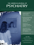
A neurological examination revealed that the patient had bilateral ptosis, infranuclear ophthalmoparesis, and generalized areflexia. Her diagnostic work-up indicated that lactate was increased two-fold above the upper limit of normal in the serum and CSF. Her urine analysis for purines and pyrimidines yielded increased concentrations of thymidine and deoxyuridine. Electromyography showed marked demyelinating sensorimotor polyneuropathy, and diffuse leukoencephalopathy was found by MRI ( Figure 1 ). A genetic analysis revealed a hitherto nondescribed homozygous mutation (c.605G>A,p.Arg202Lys) in the ECGF1 gene, which encodes for thymidine phosphorylase. This mutation could not be detected in 68 healthy, ethnically matched comparison subjects from India. Miss A was then diagnosed as having mitochondrial neurogastrointestinal encephalomyopathy.

a The magnetic resonance images illustrate increased signal intensity (arrowheads). The images demonstrate diffuse leukoencephalopathy with involvement of the A) centrum semiovale (axial T2-weighted images), B) parts of the thalamus (axial T2-weighted images), C) internal capsule (coronal fluid-attenuated inversion recovery images), and D) pons (sagittal T2-weighted images).
With regard to the present case of prominent cachexia in mitochondrial neurogastrointestinal encephalomyopathy mimicking anorexia nervosa, we would like to emphasize that syndromes of eating disturbances and cachexia could be the most prominent features of a number of somatic and psychiatric disorders. In addition to prominent eating disturbance in common psychiatric diseases such as anorexia nervosa, depression, and schizophrenia, such behavior and symptoms can also be prominent in mainly somatic disorders such as juvenile Alexander disease, Klinefelter’s syndrome, hypothalamic brain tumors, Lyme disease, Crohn’s disease, celiac disease, achalasia, and Luft’s disease.
Thus, thorough clinical (particularly neurological) and biochemical examinations (including CSF and urine analysis) together with MRI are essential to clearly differentiate between psychiatric and somatic origins within the spectrum of cachexia inducing disorders, especially in cases of atypical anorexia nervosa, which lack cardinal symptoms (distorted body image, intentional weight loss), and in cases in which abdominal complaints may be prominent.
Our patient eventually received better absorbable high-caloric nutrition via a gastric tube in addition to intravenous feeding. These measures resulted in a weight gain to 28.6 kg (body mass index: 12.7) and improvement of her physical state, which remained stable 6 months following her discharge from the hospital.
1. Lara MC, Valentino ML, Torres-Torronteras J, Hirano M, Martí R: Mitochondrial neurogastrointestinal encephalomyopathy (MNGIE): biochemical features and therapeutic approaches. Biosci Rep 2007; 27:151–163Google Scholar
2. Hirano M, Lagier-Tourenne C, Valentino ML, Marti R, Nishigaki Y: Thymidine phosphorylase mutations cause instability of mitochondrial DNA. Gene 2005; 354:152–156Google Scholar