
Structure of the Human Prefrontal Cortex
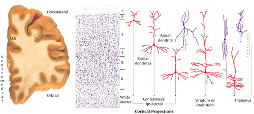
The prefrontal cortex refers to a structurally and functionally heterogenous group of cortical areas located anterior to the motor and premotor regions of the frontal lobe. As shown in an unstained coronal block (left panel), cut immediately anterior to the corpus callosum through the left hemisphere of a postmortem human brain, the prefrontal cortex is a several millimeter thick ribbon of gray matter (appearing light brown in this image) that can be divided into dorsolateral, orbital, and ventromedial regions. This block also includes the adjacent anterior cingulate gyrus (ACG) of the limbic lobe. Each of these regions differs in cytoarchitecture, that is, the size and packing density of the constituent neurons. A Nissl-stained section (center panel) from the dorsolateral prefrontal cortex (blue box in left panel) shows the typical arrangement of cortical neurons into six layers or lamina, numbered from the pial surface of the cortex to the underlying white matter. Distributed across these layers are different types of pyramidal neurons (red cells in right panel), representing about 75% of cortical neurons, which typically have triangularly-shaped cell bodies, a single apical dendrite directed toward the pial surface, and an array of basilar dendrites. Depending on their laminar location, the axons of pyramidal neurons provide excitatory projections to different brain regions, with those located in layers 2-3 projecting principally to other cortical regions in the same (ipsilateral) or opposite (contralateral) hemisphere, those located in layer 5 projecting principally to the striatum or brainstem, and those in layer 6 projecting principally to the thalamus. Axons that project to the prefrontal cortex from other brain regions also tend to innervate different subsets of cortical layers. For example, axonal projections (green) from the thalamus terminate in layers deep 3 and 4. The remaining 25% of prefrontal neurons are local circuit or interneurons (blue cells). These neurons use the inhibitory neurotransmitter GABA and have axons that arborize locally and innervate other neurons in the same area of the prefrontal cortex.
Address reprint requests to Dr. Tamminga, UT Southwestern Medical Center, Department of Psychiatry, 5323 Harry Hines Blvd., #NC5.914, Dallas, TX 75390-9070; [email protected] (e-mail).
prefrontal cortex

