Neural Networks
Advances in human brain imaging methods have provided a remarkable opportunity for studying the neural basis of human brain function. Usually the goal of these studies is the localization of specific task components to defined brain areas. More recent efforts have attempted to identify functional neural networks in the central nervous system. Still, with either approach, there exists the question of how regional cerebral blood flow (rCBF) data reflect underlying neuronal events such as those measured by electrophysiological recording (e.g., cell firing), since imaging data are an indirect measure of neuronal activity. It is generally thought that rCBF mainly reflects synaptic activity rather than neuronal firing per se. Synaptic activity can lead to an increase in both rCBF and brain oxidative metabolism, whether that activity results in inhibition or excitation. Therefore, both excitatory and inhibitory synaptic activity may increase rCBF even if the inhibition causes a decrease in overall local or distant neuronal firing rates.
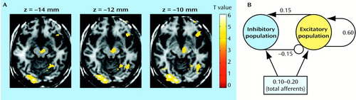
We have used computational modeling to examine factors that potentially play a role in the relationship between neuronal events and the signals measured in human imaging experiments. The goal of computational modeling is to develop a biologically plausible mathematical model that can simulate human behavior in simple behavioral tasks. Consistently, experimental studies indicate that there is a close correlation between neuronal excitation and functional imaging measures such as rCBF. Human imaging studies, like the functional magnetic resonance imaging (MRI) activation map of visual discrimination seen in part A of the figure above, can be simulated by presenting a series of stimuli to a model network (seen in part B of the figure) and spatially and temporally summing the synaptic activity of the model. The parameters of the model are chosen to represent a basic element such as a cortical column. Groups of basic elements are then combined into local regions, and finally interconnecting regions create the full network. The summation of the synaptic activity across the model should match the functional MRI map if the neuronal model of the behavior is correct. The model can then be “lesioned” to characterize a pathological process, and the resulting changes in the behavior of the network can be observed and compared to functional images that reflect the same pathological process.
Address reprint requests to Dr. Tamminga, Maryland Psychiatric Research Center, University of Maryland, P.O. Box 21247, Baltimore, MD 21228. Image is courtesy of Dr. Medoff.
figure
On the left (part A), a functional MRI activation map depicts a single individual performing a visual recognition task. On the right (part B) is the computational model representation of the local circuit involved in the task. Arrows depict excitatory synaptic activity; inhibitory synaptic activity is depicted by an open circle. The connection weights of the basic circuit are based on experimental data: 60% of all connections are local and excitatory-to-excitatory, 15% are local and excitatory-to-inhibitory, 15% are local and inhibitory-to-excitatory, and the remaining 10% are from other areas.