Fetal and Neonatal Brain Development
The effect of maternal psychiatric medication on fetal health during pregnancy is a question that lacks a precise answer or even much relevant data as yet. While clinicians discourage the use of CNS-active medications during pregnancy, there are situations where this is not possible. An illustration of this kind of clinical situation is detailed in this month’s Clinical Case Conference by Yaeger and colleagues (p. 2064). Experiments in animals looking at the effect of maternal medications on fetal CNS are only now beginning. However, and more important, the examination of this question in human neonates has already begun. Structural brain changes associated with psychosis and other major psychiatric illnesses are thought to develop early, during fetal or neonatal life. Therefore, ways of assessing structural parameters of early brain development might contribute to assessing mental health in the fetus and neonate. We have previously shown that three-dimensional ultrasound can provide reliable measures of lateral ventricular volume comparable to MRI. However, structural images of neonates, captured without sedation, are more informative and illustrate not only the structural characteristics of the brain, but also their changes during early development ( top row of MRI images ).

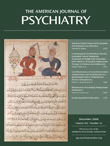
Figure 1. In the top row of longitudinal T1-weighted magnetic resonance images of the same child (and same scale), note the dramatic increase in total brain size as well as in white matter intensity over early development. In the bottom row of diffusion tensor images, white matter tractography of a neonate, one year old, and adult shows the organization of corpus callosum white matter fibers, reflected in increasing fractional anisotropy (yellows, reds), developing with age.
Most white matter in neonates is unmyelinated and therefore exhibits a low intensity on T1-weighted images relative to gray matter. Myelination occurs rapidly in the first year of life and overall adult patterns are present at age 2 years. The asymmetric (left>right) ventricular volume seen in older children is also true for neonates, suggesting that these asymmetries are present from birth. Diffusion tensor imaging allows the representation of developing white matter fiber tracts. As seen in the bottom row of images, the fractional anisotropy of white matter fibers in the corpus callosum increases with age, reflecting increasing organization and myelination. The white matter of the corpus callosum undergoes considerable maturation beginning just after birth, potentially representing a window of vulnerability for perinatal insults. The application of these brain imaging approaches to the examination of fetal and neonatal brain development could provide valuable information on pregnancy management as well as the neurodevelopmental origins of psychiatric illness.