Cognition
Visual information entering the eye undergoes a series of remarkable transformations from retina to visual thalamus to visual cortex, yielding visual perception by mechanisms that we are only beginning to understand. One of these transformations is the separation of visual information into channels, or pathways, that process different aspects of vision, including color, form, depth, and motion. This pattern of separation and specialization has been demonstrated physiologically, in the response properties of single neurons and groups of neurons, and anatomically, both in the organization of these neurons into compartments within visual cortex and in the pattern of connections that these compartments make. Indeed, this strikingly high degree of functional organization in visual cortex has provided important clues about the computational strategy the visual system employs for visual information processing. For example, the extraction of form information begins as the determination of the orientation angle (e.g., vertical, horizontal) of the edges of a visual object. In primary visual cortex, V1, the Nobel prize-winning studies of Hubel and Wiesel demonstrated how neurons processing a particular visual feature, such as orientation, are systematically laid out according to both a complete range of the various orientations, from 0 to 180 degrees, and retinotopy, the location of the object in the visual field. Furthermore, other visual properties, such as eye preference (ocular dominance), are also represented within the same cortical area in an interlaced fashion. This arrangement neatly facilitates the processing of multiple aspects or dimensions of visual information for any given point in visual space.
Beyond V1 are a number of extrastriate visual areas that also each have a high degree of functional organization. The specific set of visual properties that are organized changes from visual area to area; these differences are important indications of the progression of visual information processing as one ascends the visual hierarchy. However, the basic principles of an interlaced layout of several distinct visual properties are retained. In the first extrastriate area, V2, properties such as retinotopy, color, and orientation are also arranged systematically but in larger structures known as the V2 stripes. Seemingly, the prominent ocular dominance organization in V1 has been traded for a well-developed organization in V2 for binocular interaction (retinal disparity, which yields stereoscopic depth). Whether the purpose of the extensive pattern of functional organization and compartmentalization in these visual areas is merely for developmental convenience or perhaps for the facilitation of forming specific connections and computations on an ongoing basis, their existence must reflect the processing strategies employed and the overall role of that visual area in visual perception.
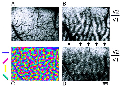
Functional organization of visual areas V1 and V2as revealed by the method of optical imaging of neuronal activity. A: An image of the surface and vasculature of a 9-mm-by-6-mm portion of primate visual cortex. B: The optical imaging map of ocular dominance from the same portion of visual cortex. The dark bands represent columns dominated by the left eye and the light bands, the right eye. Note the absence of ocular dominance structure in the imaged strip of V2 (topmost 1.5 mm). C: A map of orientation tuning produced by a vector combination of frames acquired during stimulation with four orientations (coded in color, as shown). D: Image of activity (darkening) because of left eye stimulation as compared to the no-stimulus condition (complementary right eye image not shown).
Figure reproduced by permission of the American Association for the Advancement of Science (Ts’o et al., Science 1990; 249:418).