Brain Development, III: Cerebral Cortex
Development of the adult cerebral cortex is determined by several distinct processes (genetic, molecular, and activity-dependent), which together produce the extensive functional specification characteristic of the cortex. In humans, the cerebral cortex must have not only broad functional capacity but also the ability to adapt for change in function and to provide new functional domains as needed. Early regionalization of the neural tube is extensively driven by genetic signals (see “Brain Development, I,” March 1998 issue). The fetal ventricular zone in the cerebral wall supplies the cells for cerebral cortical development. In the ventricular zone exists a pool of stem cells (self-renewing) and progenitor cells (restricted potential). The progenitor cell pool initially expands, then undergoes neurogenesis; these neurons subsequently undergo directed radial migration (perpendicular to the cortical surface) to populate the cerebral cortex. At the progenitor-pool phase, the progenitor cells can mix among other cells of the cortical pool but cannot cross between the striatal and cortical segments. There is an early phenotypic identification of the progenitor cells (while in the ventricular zone) to cortical area and lamina probably related to unique contributions of gene expression and to local environmental signals. After final cellular mitosis in the ventricular region, radial migration of neurons occurs in an inside-out manner. The period of cell fate specification occurs when the progenitor cells still reside in the ventricular zone and is probably early, short, and critical to ultimate cortical development. Although early specification of cell fate occurs (figure), many mechanisms for late cortical plasticity exist; these are extensively driven by extrinsic sensory inputs to the cortex. This activity-dependent phase of development is particularly important for establishing precise functional cortical maps for the efficient performance of higher mental tasks.
Address reprint requests to Dr. Tamminga, Maryland Psychiatric Research Center, P.O. Box 21247, Baltimore, MD 21228. Figure courtesy of Dr. Roberts.

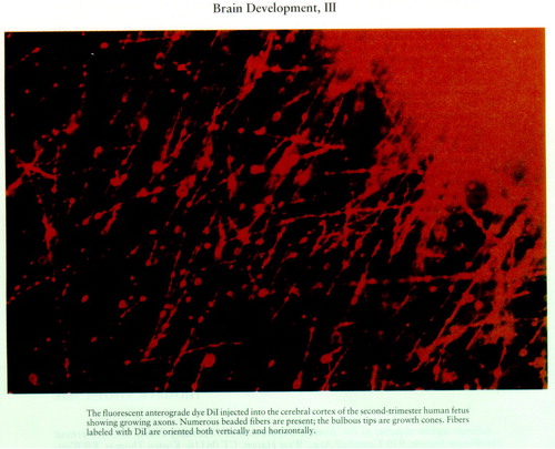
FIGURE 1. The fluorescent anterograde dye DiI injected into the cerebral cortex of the second-trimester human fetus showing growing axons. Numerous beaded fibers are present; the bulbous tips are growth cones. Fibers labeled with DiI are oriented both vertically and horizontally.


