Amygdala-Guided Neurofeedback for Major Depression
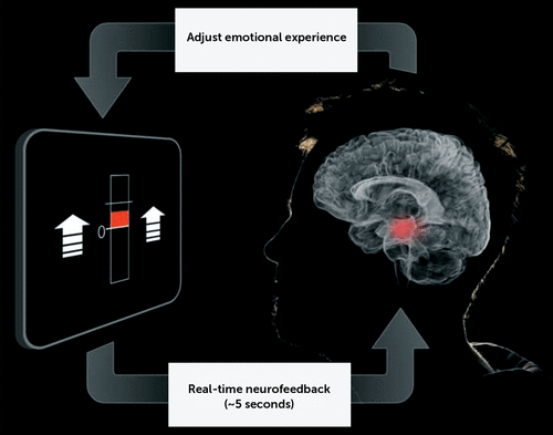
As we ride the crest of the wave in neuroscience-informed precision psychiatry, we build the foundation for novel neuroscience-informed treatment strategies. In this issue, Young et al. (1) report on the use of amygdala-focused functional MRI (fMRI) neurofeedback for alleviating emotional symptoms of major depression (Figure 1 illustrates the conceptual framework for such amygdala-guided neurofeedback). This trial is a significant proof of concept for advancing treatment strategies focused on neural substrates. Neurofeedback has a potential benefit complementary to other external neurostimulation techniques. It fosters self-regulation of the patient’s cognitive bias toward negative and positive emotions and thus increases experiences of self-efficacy, which may be important for therapeutic efficacy and treatment adherence.

FIGURE 1. Illustration of the Conceptual Framework Underlying Amygdala-Guided Neurofeedback
fMRI neurofeedback also offers the potential for neural source localization accuracy and access to deep brain structures. Deep brain stimulation also offers these advantages. Direct comparison of these techniques, and the precise way to target their application for the right patient at the right time, will be an important direction for future research. We do not yet know how fMRI neurofeedback works, and for which patients it will work. fMRI neurofeedback may foster self-regulation by correcting a primary dysfunction, such as hyper- or hypoactivation of specific brain areas or circuits. Another possibility is that it acts indirectly by activating or suppressing neural circuits that are not the primary dysfunction but that neuromodulate the dysfunctional circuit and thereby produce clinical benefits.
Young and colleagues’ design hypothesizes the first principle, the presumption of direct correction of a primary dysfunction. This design builds on an earlier pilot study (2). The focus is on a primary dysfunction of the amygdala. Amygdala activation is probed by a positive autobiographical memory paradigm. The authors’ focus on the amygdala was based on prior knowledge concerning the amygdala’s role in emotional function (e.g., 3), the pathophysiology of depression (e.g., 4), and response to antidepressants (e.g., 5). The amygdala has also been implicated in prediction of antidepressant treatment outcomes (e.g., 6).
Neurofeedback therapy consisted of asking the patients to retrieve positive memories of themselves and to increase the activation of their amygdala while doing so, presumably by focusing on their most positive memories. The patients can see the increased activation of their amygdala, determined by fMRI as increases in a blue bar. The paradigm was undertaken at a baseline session and 1 week later, and symptom outcomes were assessed 5–7 days after that. The primary outcome measure was remission of symptoms, defined as a score <10 on the Montgomery-Åsberg Depression Rating Scale (MADRS). Symptoms were also assessed using the Hamilton Depression Rating Scale, the Snaith-Hamilton Pleasure Scale, the Hamilton Anxiety Rating Scale, and the Beck Depression Inventory. Behavioral measures of autobiographical memory were recorded.
Based on this design, symptom change was assessed over a maximum period of 2 weeks after the initiation of therapy. From the initial 352 potential participants screened for the study, 36 adults with depression were randomly assigned to either amygdala-targeted neurofeedback (N=19) or a control condition targeting the parietal region (N=17). The parietal sulcus was chosen as the control region because of its putative noninvolvement in emotion regulation. The final sample for analysis comprised 18 participants in the experimental condition and 15 in the control condition. In the experimental condition, amygdala activation increased more from baseline than it did in the group assigned to parietal region control, and 33.3% of the group were defined as remitters at follow-up, compared with 6.7% of the control group. Based on criteria for response (a reduction >50% in MADRS score), 66.7% of the experimental group responded, compared with 13.3% of the control group.
How far does this study take us, and what are the next steps? The difference in the number of remitters as a function of the brain region activated by neurofeedback is promising evidence for the utility of this new brain dysfunction–guided approach. The findings generate important new lines of inquiry and the need for validation and prospective study evaluations.
This preliminary trial is highly interesting because it demonstrates a new strategy for connecting informative neural substrates with targeted clinical interventions. There are several methodological approaches that may not be feasible in an initial study, but that could be used in the future to explore the generalizability and specificity of the findings. For example, it would be helpful to understand whether particular clinical characteristics identify the patients who meet inclusion criteria for neurofeedback in the scanner, and whether the findings are specific when there is a strict as well as a “warm” placebo control that activates another region, such as portions of the frontal cortex that may be more involved in emotional regulation than the parietal cortex.
Important future directions for understanding the mechanism of action will be to consider whether neuromodulation is acting on a primary amygdala dysfunction or via interaction with other neural circuit dysfunctions. Understanding these connections will help flesh out a prospective model for more precisely guiding interventions according to the needs of the individual patient. A specific next step would be to determine which type of pretreatment neurobiological dysfunction may be a necessary precondition (or not) for the therapeutic efficacy of neurofeedback. For instance, we do not know whether the patients in this study had hypo- or hyperactivation of the amygdala relative to healthy controls at baseline. To make another step toward a mechanistic understanding of this finding, it would also be interesting to know whether neurofeedback modulation of the amygdala recruits other regions that have direct connections with the amygdala, and whether modulation is also influenced by indirect effects from other neural circuits. Clinically, because neurofeedback has the potential to foster self-efficacy and self-regulation, it would be valuable to know whether changes in amygdala activity persist beyond the 2-week follow-up period in the study, as well as whether they track ongoing clinical recovery.
1 : Randomized clinical trial of real-time fMRI amygdala neurofeedback for major depressive disorder: effects on symptoms and autobiographical memory recall. Am J Psychiatry 2017; 174:748–755Link, Google Scholar
2 : Real-time fMRI neurofeedback training of amygdala activity in patients with major depressive disorder. PLoS One 2014; 9:e88785Crossref, Medline, Google Scholar
3 : Cognitive and neural mechanisms of emotional memory. Trends Cogn Sci 2001; 5:394–400Crossref, Medline, Google Scholar
4 : Functional neuroimaging studies of the amygdala in depression. Semin Clin Neuropsychiatry 2002; 7:234–242Crossref, Medline, Google Scholar
5 : Relationship between amygdala responses to masked faces and mood state and treatment in major depressive disorder. Arch Gen Psychiatry 2010; 67:1128–1138Crossref, Medline, Google Scholar
6 : Amygdala reactivity to emotional faces in the prediction of general and medication-specific responses to antidepressant treatment in the randomized iSPOT-D trial. Neuropsychopharmacology 2015; 40:2398–2408Crossref, Medline, Google Scholar


