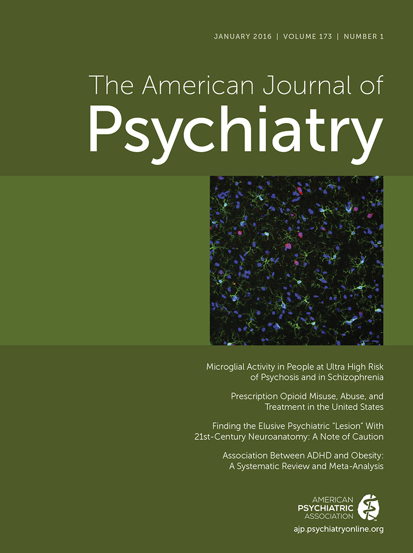
Microglial Activation and the Onset of Psychosis
Microglial activation may represent a proximal mechanism by which both immunologic and neuroplasticity-related etiological factors influence the pathophysiology of schizophrenia. Microglia are essentially immune cells operating in the brain; their activation is a key constituent of neuroinflammation. This role potentially links microglial activation with evidence of inherited structural genetic variations in and altered expression of genes that control immunologic/inflammatory signaling in schizophrenia (1). In addition, activated microglia eliminate inactive or weak synapses, a process that depends critically on long-term potentiation and other aspects of synaptic plasticity (2). This role potentially links microglial activation with evidence of inherited structural variations in and altered expression of genes involved in N-methyl-d-aspartate (NMDA)-dependent plasticity in schizophrenia (3). Whether due to disturbances in immunologic and/or plasticity-related signaling, over-active microglia may contribute to the pathophysiology of this disorder by decreasing spine and synapse density on glutamatergic cortical pyramidal neurons (4) below a threshold critical for integrated functioning of networks relevant to reality testing (5).
Some, though not all, positron emission tomography (PET) studies of the 18kD translocator protein (TSPO), which is expressed in microglia, have reported increased microglial activation in patients with schizophrenia compared with healthy control subjects (6). However, studies of patients with established illness cannot differentiate which neural changes are causal, which are epiphenomena, and which are secondary to factors associated with chronicity of illness or antipsychotic drug treatment. Teasing these alternatives apart requires prospective evaluation prior to the onset of illness and initiation of treatment.
It is thus provocative that in this issue of the Journal, Bloomfield et al. (6) report on the results of a PET study with [11C]PBR28 comparing microglial activation in a sample of individuals at ultra high risk for psychosis compared with demographically similar healthy control subjects. The binding ratio in prefrontal and temporal gray matter (relative to whole brain) was elevated in the ultra-high-risk subjects, with a positive correlation between binding ratio and prodromal symptom severity. This study is the first using a central measure of microglial activation to find evidence supporting the theory that a neuroinflammatory process may be involved in the development of psychosis. That this effect was independent of antipsychotic drug exposure in the ultra-high-risk cases (who had none) and that even larger elevations were observed in a parallel case-control study among patients with established schizophrenia are also encouraging to this view.
Because of the relative quantitation method used in this study (whereby binding potential in cortical gray matter areas was expressed as a ratio of binding potential in the brain overall), it remains ambiguous whether the higher binding ratios in ultra-high-risk cases are due to higher TSPO expression in cortex, lower TSPO expression elsewhere (e.g., white matter, subcortical nuclei), or both. When quantified using more traditional methods (volume of distribution or VT), the groups did not differ in TSPO expression in gray matter or elsewhere (6). Clearly, additional (and larger) studies of ultra-high-risk cases are needed to resolve this question.
Another limitation of the study is the lack of information on outcome among the ultra-high-risk cases; thus, we do not yet know if microglial activation specifically precedes and predicts onset of psychosis or is characteristic of at-risk mental states in general. That [11C]PBR28 binding correlates with prodromal symptom severity helps in this regard because higher prodromal symptom severity at baseline is highly predictive of conversion to psychosis in this population (7).
The search for a psychosis-triggering mechanism profits greatly from a prospective vantage point, as afforded by the prodromal risk paradigm. However, with only a single scan collected at a baseline assessment, it is not clear whether microglial activation increases as cases move from an at-risk vulnerability state to a fully psychotic state, as one would expect if it participates causally in symptom formation. Longitudinal evaluation of ultra-high-risk cases can help to differentiate the likely role of a given process in pathophysiology. A marker that is stably deviant in those who develop psychosis from pre- to postonset may be necessary, but clearly is not sufficient for psychotic symptom formation. Conversely, a marker that worsens as symptoms worsen in the ramp-up to full psychosis has the potential to represent a proximal sufficient cause of psychosis (5).
If a disruption in brain connectivity, driven primarily by a reduction in dendritic spines expressing NMDA synapses that accelerates during late adolescence and early adulthood (4), is a key triggering mechanism in the onset of psychosis, we should expect to see evidence of differential change in neuroimaging markers of these processes among those who develop fully psychotic symptoms. One such marker may be an accelerated loss of gray matter among ultra-high-risk cases who convert to psychosis compared with those who do not and healthy controls (8). That this differential loss of gray matter, maximal in superior and medial prefrontal cortex, is predicted by higher levels of proinflammatory cytokines (i.e., activators of the M1 cytotoxic phenotype of microglia [9]) at baseline supports the hypothesis that increasing microglial activation may drive progressive gray matter change during the development of schizophrenia (8).
Progress on elucidating mechanisms of onset of psychosis could yield novel or repurposed compounds with more than palliative efficacy; that is, capable of preventing or mitigating the changes in brain structure and function underlying functional decline and onset of full symptoms (10). Such compounds, combined with psychosocial interventions, could also help to redirect a young person otherwise predisposed to schizophrenia toward a trajectory of social engagement, educational completion, and independent living. Linking this line of argument to the issues raised above, a question of major importance is whether increasing neuroinflammation precedes and predicts changes in cortical gray matter and functional connectivity prior to onset. The answer is critical; if yes, a prevention trial with anti-inflammatory agents would be strongly indicated, but if no, clearly not. In another possible cascade, dysregulated NMDA-dependent synaptic plasticity may be a proximal cause of the accelerated gray matter decline and disrupted functional connectivity seen in those who convert to psychosis (via pruning of overabundant weak synapses), with neuroinflammation (microglial activation) representing a secondary signal (i.e., as in a clean-up operation). In this case, targeting NMDA-dependent plasticity mechanisms would be indicated over other potential strategies. Parsing these alternatives will require research designs involving multiple points of observation prior to onset of psychosis and application of time-lagged and growth curve analytic approaches that can help to establish temporal precedence and mediation among multiple cascading influences over time. Alternatively, we may find evidence of feed-forward mediation of psychosis risk primarily through inflammatory processes in some patients and plasticity-related processes in others, in which case, biomarkers may be useful for selecting interventions targeted to particular mechanisms for different individual patients (5).
1 : Inflammation and immunity in schizophrenia: implications for pathophysiology and treatment. Lancet Psychiatry 2015; 2:258–270Crossref, Medline, Google Scholar
2 : The “quad-partite” synapse: microglia-synapse interactions in the developing and mature CNS. Glia 2013; 61:24–36Crossref, Medline, Google Scholar
3 : Genetic risk for schizophrenia: convergence on synaptic pathways involved in plasticity. Biol Psychiatry 2015; 77:52–58Crossref, Medline, Google Scholar
4 : Dendritic spine pathology in schizophrenia. Neuroscience 2013; 251:90–107Crossref, Medline, Google Scholar
5 : How schizophrenia develops: cognitive and brain mechanisms underlying onset of psychosis. Trends Cogn Sci (Epub ahead of print, Oct 19, 2015)Medline, Google Scholar
6 . Microglial activity in people at ultra high risk of psychosis and in schizophrenia: an [11C]PBR28 PET brain imaging study. Am J Psychiatry 2016; 173:44–52Link, Google Scholar
7 : Prediction of psychosis in youth at high clinical risk: a multisite longitudinal study in North America. Arch Gen Psychiatry 2008; 65:28–37Crossref, Medline, Google Scholar
8 : Progressive reduction in cortical thickness as psychosis develops: a multisite longitudinal neuroimaging study of youth at elevated clinical risk. Biol Psychiatry 2015; 77:147–157Crossref, Medline, Google Scholar
9 : Microglia and macrophages of the central nervous system: the contribution of microglia priming and systemic inflammation to chronic neurodegeneration. Semin Immunopathol 2013; 35:601–612Crossref, Medline, Google Scholar
10 : Cure therapeutics and strategic prevention: raising the bar for mental health research. Mol Psychiatry 2006; 11:11–17Crossref, Medline, Google Scholar