Reduced Amygdala Response in Youths With Disruptive Behavior Disorders and Psychopathic Traits: Decreased Emotional Response Versus Increased Top-Down Attention to Nonemotional Features
Abstract
Objective:
Amygdala dysfunction has been reported to exist in youths and adults with psychopathic traits. However, there has been disagreement as to whether this dysfunction reflects a primary emotional deficit or is secondary to atypical attentional control. The authors examined the validity of the contrasting predictions.
Method:
Participants were 15 children and adolescents (ages 10–17 years) with both disruptive behavior disorders and psychopathic traits and 17 healthy comparison youths. Functional MRI was used to assess the response of the amygdala and regions implicated in top-down attentional control (the dorsomedial and lateral frontal cortices) to emotional expression under conditions of high and low attentional load.
Results:
Relative to youths with disruptive behavior disorders and psychopathic traits, healthy comparison subjects showed a significantly greater increase in the typical amygdala response to fearful expressions under low relative to high attentional load conditions. There was also a selective inverse relationship between the response to fearful expressions under low attentional load and the callous-unemotional component (but not the narcissism or impulsivity component) of psychopathic traits. In contrast, the two groups did not differ in the significant recruitment of the dorsomedial and lateral frontal cortices as a function of attentional load.
Conclusions:
Youths with disruptive behavior disorders and psychopathic traits showed reduced amygdala responses to fearful expressions under low attentional load but no indications of increased recruitment of regions implicated in top-down attentional control. These findings suggest that the emotional deficit observed in youths with disruptive behavior disorders and psychopathic traits is primary and not secondary to increased top-down attention to nonemotional stimulus features.
Youths with disruptive behavior disorders, including conduct disorder and oppositional defiant disorder, show increased aggression and antisocial behavior (1). A subset of these youths also display psychopathic traits, including callous-unemotional (e.g., lack of guilt and empathy), narcissistic (e.g., excessive bragging about one's abilities), and impulsive (e.g., acting without thinking) components (2). These traits are detectable early in childhood and persist into adulthood (3). Youths with both disruptive behavior disorders and psychopathic traits are at highest risk for recurrent antisocial behavior (1, 4). However, functional MRI (fMRI) research investigating the pathophysiology of these disorders and traits has only recently begun (e.g., 5–9).
The presence of psychopathic traits, particularly the callous-unemotional component, is thought to reflect emotional processing deficits (10–12) and dysfunction in the amygdala (11, 13), ventromedial frontal cortex, and caudate (6), although other regions, such as the cingulate cortex, may also be implicated (14). It is argued that psychopathic traits reflect impairment in stimulus-reinforcement learning and decision making (13). From this perspective, a reduced amygdala response results in impairment in an individual's ability to learn to avoid actions associated with the distress of others (e.g., a victim's sadness). In line with this, youths with both disruptive behavior disorders and psychopathic traits show reduced autonomic responses to the distress of others (15), reduced recognition of fearful expressions (16), and reduced amygdala response to fearful expressions (7, 8). Moreover, reduced amygdala response to sad expressions in youths with conduct disorder has been reported (9).
However, an alternative position on psychopathic traits has been prevalent for some time (17, 18). According to this view, the emotional dysfunction is not primary but rather is a secondary consequence of an irregularity in attention (17, 18). From this standpoint, the emotional deficits should be understood as a failure to process information that is peripheral to the focus of attention (18). These problems with deliberate focusing of attention “can be understood as difficulty accommodating bottom-up, stimulus-driven information, especially when the bottom-up information is inconsistent with or unrelated to the current top-down, effortful focus of attention” (17, p. 346).
Until recently (19), this attention-based model has been focused on descriptions of information processing functions rather than their neural correlates. However, it is clear that brain regions implicated in top-down attentional control, such as the lateral frontal, dorsomedial, and parietal cortices, affect the amygdala's response to emotional stimuli. Increased priming of task-relevant representations by these regions is thought to reduce the representational strength of emotional stimuli within the temporal cortex, following representational competition (20), and consequently reduces amygdala responses to these stimuli (21, 22). In short, the reduced emotional responsiveness of individuals with elevated psychopathic traits might be a secondary consequence of heightened top-down attentional control in response to nonemotional stimulus features (23).
We tested two contrasting hypotheses regarding the basis of emotion dysfunction in youths with disruptive behavior disorders and psychopathic traits. Our first hypothesis was that amygdala-based emotion dysfunction is primary (11, 13, 14). This predicts that 1) under low attentional load conditions, there will be a reduced amygdala response to fearful expressions in these youths, but under high attentional load conditions, both these youths and healthy comparison subjects will show a reduced amygdala response to fearful expressions, and 2) both groups will show appropriate and equivalent significant increases in activity in brain regions implicated in top-down attentional control (the dorsomedial and lateral frontal cortices) as a function of attentional load. Our secondary hypothesis was that the emotion dysfunction is secondary to attentional irregularities (17, 18, 24). This also predicts reduced amygdala responsiveness to fearful expressions under low attentional load conditions in youths with behavior disorders and psychopathic traits. However, this position predicts that these youths will show significantly increased responses, relative to healthy youths, within regions implicated in top-down attentional control under low attentional load.
Method
Participants
Of the 32 youths (ages 10–17 years) who participated in this study, 15 had disruptive behavior disorders and psychopathic traits and 17 were healthy comparison subjects. Demographic and clinical characteristics of both groups are summarized in Table 1. The youths were recruited from the community through advertising, flyers, and referrals from mental health practitioners. Participants and their parents provided informed assent and consent. This study was approved by the National Institute of Mental Health Institutional Review Board.
| Characteristic | Disruptive Behavior Disorders Plus Psychopathic Traits Group (N=15) | Healthy Comparison Group (N=17) | ||
|---|---|---|---|---|
| Mean | SD | Mean | SD | |
| Age (years) | 15.67 | 2.53 | 14.50 | 2.14 |
| IQa | 96.67 | 8.83 | 101.19 | 12.95 |
| Antisocial Process Screening Device scores | ||||
| Total | 29.13* | 5.17 | 4.47* | 3.00 |
| Callous-unemotional traits | 8.33* | 1.80 | 1.47* | 1.33 |
| Narcissistic traits | 8.87* | 3.02 | 1.12* | 1.27 |
| Impulsive traits | 7.73* | 1.75 | 1.88* | 1.17 |
| N | % | N | % | |
| Gender | ||||
| Male | 12 | 80 | 9 | 52.9 |
| Female | 3 | 20 | 8 | 47.1 |
| DSM-IV diagnosis | ||||
| Conduct disorder | 11 | 73.33 | ||
| Oppositional defiant disorder | 4 | 26.67 | ||
| Attention deficit hyperactivity disorder | 8 | 53.33 | ||
TABLE 1. Demographic and Clinical Characteristics of Youths With Disruptive Behavior Disorders and Psychopathic Traits and Healthy Comparison Subjects
The Schedule for Affective Disorders and Schizophrenia for School-Age Children (K-SADS [25]) was administered to all youths and their parents by an experienced clinician who was trained and supervised by expert child psychiatrists (interrater reliability >0.75 for all diagnoses). The Wechsler Abbreviated Scale of Intelligence (two-subtest form) was used to assess IQ. Individuals were excluded from participation if they had pervasive developmental disorder; Tourette's syndrome; a lifetime history of psychosis; depression; bipolar disorder; generalized, social, or separation anxiety disorder; posttraumatic stress disorder; a neurologic disorder; a history of head trauma; a history of substance abuse; or an IQ <80. Additionally, parents completed the Antisocial Process Screening Device (26), a measure of psychopathic traits. Youths who met K-SADS criteria for conduct disorder or oppositional defiant disorder and who had a score of 20 or greater on the Antisocial Process Screening Device were included in the behavior disorders plus psychopathic traits group; those who met criteria for conduct disorder or oppositional defiant disorder but had a score <20 were excluded from the study. Individuals included in the comparison group did not meet criteria for any diagnosis according to K-SADS and had a score <20 on the Antisocial Process Screening Device. The groups did not differ significantly in age or IQ or in terms of racial and gender breakdown (Table 1).
Study Measures
Antisocial Process Screening Device.
The Antisocial Process Screening Device is a 20-item parent-completed rating scale designed to assess three dimensions related to psychopathic traits in youths: callous-unemotional traits, narcissism, and impulsivity. There is no established cutoff score for classification of a high level of psychopathic traits (26). In line with previous work (5, 8), we used a cutoff score of 20. All healthy comparison subjects in our study had a score of 11 or lower on this measure. The measure was completed by the parents during screening prior to their children's entry into the study.
Bars task (27).
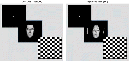
We used a modified version of the emotion-attention bars task used in previous studies (27, 28). In this task, participants were presented with faces flanked by lines (Figure 1). The faces displayed either a fearful or a neutral affect. The flanking lines were either parallel (50% of trials) or nonparallel. Attentional load was manipulated by varying the degree of deviation on nonparallel trials: 90° in the low-load (easy) condition, 24° in the medium-load condition, and 16° in the high-load (hard) condition. Participants responded to the stimuli via button press according to whether the lines were parallel or nonparallel. Trials began with the presentation of the stimulus (a face bracketed by two bars) for 200 msec, followed by a blank screen for 1800 msec and a 300-msec fixation. Participants could respond at any point during the trial.

FIGURE 1. Emotion-Attention Bars Taska
a Participants indicated via button press whether the lines displayed were parallel (50% of trials) or not. Fifty percent of trials depicted fearful expressions, and the remaining 50% of trials depicted neutral expressions.
Participants completed one brief practice run outside of the scanner and then five task runs in the scanner. Each run consisted of 128 trials (48 parallel trials; 16 low-load, 16 medium-load, and 16 high-load trials; and 32 fixation trials to provide a baseline). Fearful expressions were displayed in half of the trials, and neutral expressions were displayed in the other half. The run order and the trial order within runs were randomized across participants. Because of technical difficulties, data were available for only three runs for two participants in the behavior disorders plus psychopathic traits group, and data for only four runs were available for one individual in the healthy comparison group and one in the disorders group.
MRI Parameters
Participants were scanned using a 1.5-T GE Signa scanner (General Electric, Waukesha, Wisc.). A total of 139 functional images per run were taken, with a gradient echo planar imaging (EPI) sequence (repetition time=2500 msec; echo time=30 msec; matrix=64×64; flip angle=90°; field of view=24 cm). Whole-brain coverage was obtained using 31 axial slices (thickness=4 mm; in-plane resolution=3.75 mm × 3.75 mm). A high-resolution anatomical scan (three-dimensional spoiled gradient recalled acquisition in a steady state; repetition time=8.1 msec; echo time=1.8 msec; field of view=24 cm; flip angle=20°; 128 axial slices; thickness=1.5 mm; matrix=256×256) in register with the EPI data set was obtained; this scan covered the whole brain.
Imaging Data Preprocessing
Imaging data were preprocessed and analyzed using the Analysis of Functional NeuroImages (AFNI) software package (29). At the individual level, functional images from the first six repetitions, collected before equilibrium magnetization was reached, were discarded. Functional images from the five time series were motion-corrected and spatially smoothed with a 6-mm full-width half-maximum Gaussian filter. The time series were normalized by dividing the signal intensity of a voxel at each point by the mean signal intensity of that voxel for each run and multiplying the result by 100. Resultant regression coefficients represented a percentage of signal change from the mean.
Accordingly, the following 10 regressors were generated: neutral parallel, neutral low-load, neutral medium-load, neutral high-load, fear parallel, fear low-load, fear medium-load, fear high-load, incorrect response, and no-response trials. All regressors were created by convolving the train of stimulus events with a gamma variate hemodynamic response function to account for the slow hemodynamic response. Linear regression modeling was performed using the 10 aforementioned regressors plus regressors to model a first-order baseline drift function. This produced a beta coefficient and associated t statistic value for each voxel and regressor. In accordance with previous findings that normalization of brain volumes from age 7–8 years onward does not introduce major age-related distortions in localization or time course of the blood-oxygen-level-dependent (BOLD) signal in event-related fMRI (30, 31), the participants' anatomical scans were individually registered to the Talairach-Tournoux brain atlas (32). Functional EPI data were then registered to the Talairach anatomical scans within AFNI.
fMRI Data Analysis
The group analysis of the BOLD data was then performed with regression coefficients from individual subject analyses using a 2×2×2 (group-by-emotion-by-attentional load) whole-brain repeated-measures analysis of variance (ANOVA). Initial thresholding was set at a p value of <0.005, with an extent threshold of 10 voxels, a combination that has been demonstrated to produce a desirable balance between type I and II error rates (33). The average percentage of signal change was measured within each significant cluster of 10 or more voxels. Post hoc analyses of significant main effects and interactions were conducted using t tests in SPSS, version 19.0 (SPSS, Inc., Chicago), to further characterize the percentage of signal change. Because of concerns about the reduction in statistical power associated with a three-level analysis, only the low and high attentional load conditions were included in the ANOVA involving BOLD data.
Results
Behavioral Data Analysis
We conducted two 2×2×2 (group-by-emotion-by-attentional load) repeated-measures ANOVAs on error rate and reaction time data. The first analysis revealed a significant main effect for attentional load; participants were significantly less accurate in high- relative to low-load trials (F=88.29, df=1, 30, p<0.001). The second analysis also revealed a significant main effect for attentional load; participants were significantly faster in low- relative to high-load trials (F=54.91, df=1, 30, p<0.001). Additionally, there was a significant emotion-by-attentional load interaction (F=6.41, df=1, 30, p<0.05); participants responded significantly faster in neutral low-load trials relative to fear low-load trials (t=–2.98, df=31, p<0.01) but were equally fast in their responses in neutral high-load trials relative to fear high-load trials. There were no significant main effects or interactions involving group (see Table 1 in the data supplement accompanying the online edition of this article).
fMRI Data Analysis
Our goal was to assess potential functional irregularities within the amygdala and regions implicated in top-down attentional control in youths with behavior disorders and psychopathic traits during an emotion/attention paradigm. We examined this through a 2×2×2 repeated-measures ANOVA conducted on BOLD data (Table 2). Below we describe the key interactions with respect to our predictions (group-by-emotion-by-attentional load; group-by-emotion) and the main effects of both group and attentional load to provide the results for the test of our a priori hypotheses. No significant finding was observed for the group-by-attentional load interaction.
| Areas of Peak Activationb | Analysis | |||||
|---|---|---|---|---|---|---|
| Interaction and Regiona | Hemisphere | Brodmann's Area | x, y, z | F (df=1, 30) | p | Voxels |
| Group-by-emotion-by-attentional load | ||||||
| Amygdala/lentiform nucleusc | Left | –19.5, –13.5, –3.5 | 11.67 | 0.002 | 7 | |
| Group-by-emotion | ||||||
| Middle temporal gyrus | Left | 21 | –52.5, –1.5, –15.5 | 10.75 | 0.003 | 13 |
| Middle temporal gyrus | Left | 21 | –58.5, –7.5, –12.5 | 9.38 | 0.005 | 12 |
| Main effect of attentional load | ||||||
| Dorsomedial frontal cortex | Left | 32 | –7.5, 7.5, 41.5 | 14.47 | 6.5E-4 | 84 |
| Inferior frontal cortex/insula | Right | 45/13 | 31.5, 13.5, 8.5 | 11.56 | 0.002 | 26 |
| Inferior frontal cortex/precentral gyrus | Right | 44/13 | 49.5, –1.5, 14.5 | 9.45 | 0.005 | 25 |
| Lingual gyrus | Right | 19 | 34.5, –70.5, –3.5 | 10.69 | 0.003 | 19 |
| Main effect of group | ||||||
| Middle frontal gyrus | Right | 10 | 31.5, 49.5, 14.5 | 12.06 | 0.002 | 12 |
| Superior frontal gyrus | Right | 9 | 16.5, 46.5, 32.5 | 13.62 | 8.9E-4 | 16 |
| Superior frontal gyrus | Left | 9 | –13.5, 43.5, 29.5 | 9.91 | 0.004 | 10 |
| Inferior frontal cortex | Left | 45/47 | –37.5, 19.5, 11.5 | 16.13 | 3.4E-4 | 35 |
| Posterior cingulate cortex | Right | 23 | 13.5, –43.5, 26.5 | 15.21 | 5.0E-4 | 55 |
| Inferior temporal cortex | Left | 20 | –49.5, –10.5, –21.5 | 15.20 | 5.0E-4 | 10 |
| Declive | Left | –25.5, –55.5, –21.5 | 15.01 | 5.4E-4 | 14 | |
| Lingual gyrus | Left | 18 | 13.5, –79.5, –3.5 | 10.87 | 0.003 | 26 |
TABLE 2. Brain Regions Demonstrating Differential BOLD Responses in Task Performance Among Youths With Disruptive Behavior Disorders and Psychopathic Traits and Healthy Comparison Subjects
Group-by-emotion-by-attentional load interaction.
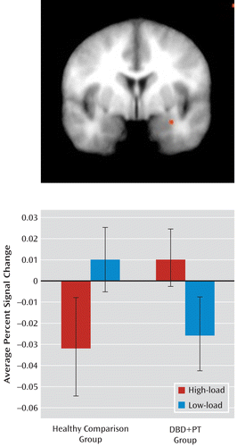
A significant three-way interaction was observed in the left amygdala/lentiform nucleus (Figure 2). Consistent with our a priori hypotheses, the healthy comparison group, relative to the disorders group, showed a significantly greater increase in amygdala response in low-load fear trials relative to high-load fear trials (t=2.42, df=30, p<0.05). Moreover, comparison subjects also showed significantly greater amygdala response to fearful expressions relative to neutral expressions compared with the disorders group only under low-load conditions (t=1.97, df=30, p<0.05). Furthermore, healthy youths, considered alone, showed significantly greater amygdala responses to fearful expressions under conditions of low relative to high attentional load (t=1.92, df=16, p<0.05, one-tailed). In contrast, youths with behavior disorders and psychopathic traits did not show significantly greater responses to fearful expressions under low relative to high attentional load.

FIGURE 2. Group-by-Emotion-by-Attentional Load Interaction in the Left Amygdala/Lentiform Nucleus in Youths With Disruptive Behavior Disorders Plus Psychopathic Traits (DBD+PT; N=15) and Healthy Comparison Youths (N=17)a
a The fMRI scan (top) illustrates significantly greater amygdala responses among healthy comparison subjects to fearful expressions under conditions of low relative to high attentional load (t=1.92, df=16, p<0.05) (the DBD+PT group did not differ significantly by condition). The healthy comparison group, relative to the DBD+PT group, also showed a significantly greater increase in amygdala response in low-load fear trials relative to high-load fear trials (t=2.42, df=30, p<0.05).
Group-by-emotion interaction.
Regions showing a group-by-emotion interaction included two areas in the left middle temporal cortex. In both these regions, no significant differences in BOLD responses to neutral expressions manifested between the study groups. However, for fearful expressions, healthy comparison subjects showed significantly greater activation than youths with behavior disorders and psychopathic traits (t=2.92, df=30, p<0.01; t=2.178, df=30, p<0.05).
Main effect of attentional load.
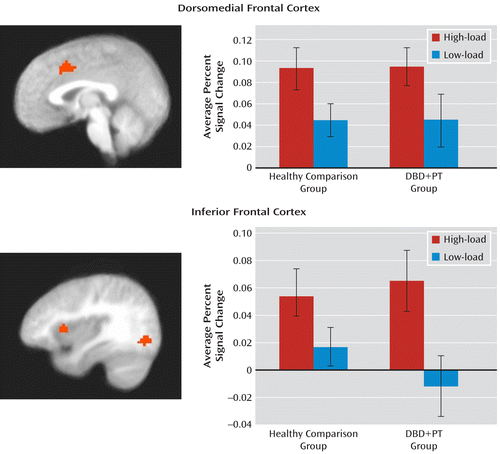
As already noted, there were no regions showing a significant group-by-attentional load interaction. However, there were several regions demonstrating a significant main effect for attentional load (Figure 3). These included the left dorsomedial frontal cortex and right inferior frontal cortex/insula. These regions showed significantly greater responses during high relative to low attentional load conditions.

FIGURE 3. Main Effect of Attentional Load in the Left Dorsomedial Frontal Cortex and Right Inferior Frontal Cortex in Youths With Disruptive Behavior Disorders Plus Psychopathic Traits (DBD+PT; N=15) and Healthy Comparison Youths (N=17)a
a The fMRI scans illustrate significantly greater activation in the left dorsomedial frontal cortex and right inferior frontal cortex in high attentional load conditions relative to low attentional load conditions, which was observed in all participants (no differences were observed between the healthy comparison and DBD+PT groups).
Main effect of group.
Brain regions showing a main effect for group included the right middle frontal gyrus, the left and right superior frontal gyrus, the left inferior frontal gyrus, the right posterior cingulate cortex, and the left inferior temporal cortex. In all of these regions, healthy youths showed greater BOLD responses than youths with behavior disorders and psychopathic traits.
Potential confounds.
To account for possible effects of medication use on BOLD responses, the preceding analysis was repeated excluding the four youths in the disorders group who were receiving medication treatment. The effects of interest were replicated with proximal activations in the same brain regions for each main effect and interaction, including a significant group-by-emotion-by-attentional load interaction within the left amygdala/lentiform nucleus (Montreal Neurological Institute [MNI] coordinates: –27, –5, –13). The only exception was for the main effect of group, in which the result for the left inferior temporal cortex was significant only at a more lenient threshold (p<0.05).
To account for possible effects of comorbid attention deficit hyperactivity disorder (ADHD), the analysis was also repeated excluding the eight youths in the disorders group who met criteria for ADHD. Again, the preceding effects of interest were replicated with proximal activations in the same brain regions for each effect and interaction. Indeed, the significant group-by-emotion-by-attentional load interaction was observed to be bilateral (left amygdala: p<0.05, MNI coordinates: –23, –4, –11; right amygdala: p<0.005, MNI coordinates: 17, –4, –11). However, the regions showing a main effect of group were only observed at more lenient thresholds (right middle frontal gyrus: p=0.01; right middle frontal, posterior cingulate, left superior frontal, and inferior temporal cortices: p<0.03).
Symptom severity and amygdala response.
Considering the significant group differences in the responses to fearful expressions under low attentional load, we investigated whether there was a relationship between reduced emotional response to fearful expressions and severity of specific components of psychopathic traits (i.e., callous-unemotional, narcissism, or impulsivity). This analysis revealed a significant inverse relationship between the amygdala responses to fearful relative to neutral expressions and scores on the callous-unemotional subscale (r=–0.376, p<0.05) of the Antisocial Process Screening Device.
Discussion
This study examined whether reduced neural response to emotional stimuli in youths with disruptive behavior disorders and psychopathic traits only occurs in the context of heightened recruitment of top-down attentional systems. As expected (21, 22, 28), healthy comparison youths showed significantly reduced amygdala response to fearful expressions in high relative to low attentional load conditions. In contrast, youths with behavior disorders and psychopathic traits did not. Indeed, the healthy youths showed a significantly greater decrease in amygdala response in high- relative to low-load fear trials compared with youths in the disorders group. Furthermore, symptom severity, as indexed by the callous-unemotional component of psychopathic traits (but not the narcissistic or impulsive component), was found to be significantly correlated with the amygdala response to fearful expressions under low attentional load. Finally, and consistent with previous findings (21, 22, 28), regions associated with top-down attentional control (the dorsomedial and lateral [inferior] frontal cortices) increased in activation with increased attentional load. Critically, both groups showed equivalent levels of recruitment of these regions with increased attentional load.
Previous studies have reported that youths with disruptive behavior disorders and psychopathic traits show reduced amygdala responses to fearful (7, 8) and sad (9) expressions. However, those studies assessed responses to emotional expressions only while participants distinguished the gender of the individual face depicted. From the perspective of attention-based accounts, the previous results could reflect a secondary consequence of heightened processing of the stimulus features relating to gender at the expense of those associated with emotion. Notably, the previous studies did not employ experimental conditions whereby attention was manipulated, and thus they could not systematically address the issue.
Our study echoes previous work (7, 8) and, critically, demonstrates that youths with behavior disorders and psychopathic traits, compared with healthy youths, show significantly less increase in the amygdala response to fearful expressions under low relative to high attentional load. Moreover, the amygdala response to fearful expressions under low attentional load conditions was negatively associated with symptom severity as indexed by scores on the callous-unemotional subscale of the Antisocial Process Screening Device. According to the attention-based model, this could reflect heightened top-down attentional priming of task-demand stimulus features (the orientation of the bars) and, consequently, reduced representation of emotional features following representational competition (20) and thus a reduced amygdala response. If this were the case, we would expect to see group differences in the recruitment of regions implicated in top-down attention (the dorsomedial and lateral frontal and parietal cortices) (20, 34, 35), particularly under low-load conditions. However, this was not observed. Specifically, no increased recruitment of either the dorsomedial or the lateral (inferior) frontal cortex was seen in youths with behavior disorders and psychopathic traits under low-load conditions. In short, our data support the suggestion that the emotional deficit in individuals with behavior disorders and psychopathic traits is primary rather than secondary to increased top-down attention. (The main effect of task load also identified activity in the precentral and lingual gyri. This activity likely reflects increased response planning and selection [precentral gyrus] and increased top-down modulation of posterior visual processing streams [lingual gyrus] [36].)
It is interesting to note that the inverse relationship between amygdala activity in response to fearful expressions under low attentional load and symptom severity was significant only for the callous-unemotional component of psychopathic traits. Callous-unemotional traits have been previously associated with amygdala dysfunction (37). Moreover, high callous-unemotional traits have been considered a critical dimension in the development of psychopathy (38), even though other factors may contribute to heightened scores for the narcissism and impulsivity components of the disorder (13, 38). Notably, previous research has also particularly associated callous-unemotional traits, as opposed to impulsive antisociality or narcissism, with the primary emotional deficit (38, 39). In short, our data support the suggestion that amygdala dysfunction causes the compromised emotional responding (callous-unemotional traits) that represents one developmental route to conduct disorder (13, 38).
There are five caveats that should be considered with respect to the present data. First, although there were high rates of comorbid ADHD within the behavior disorders plus psychopathic traits group, we did not include an ADHD comparison group. This was because previous studies have indicated that youths with ADHD do not present with the pathophysiology found in youths with behavior disorders and psychopathic traits (5, 8, 40). Indeed, recent work conducted by Posner et al. (40) found that youths with ADHD showed increased amygdala activation in response to fearful faces. Moreover, and mitigating this limitation, it is important to note that our subsequent group analysis excluding those youths with behavior disorders and psychopathic traits with comorbid ADHD revealed similar results, identifying proximal activations for the interactions and main effects. Second, the medications of four youths with behavior disorders and psychopathic traits could not be withheld at the time of scanning. However, mitigating this limitation, the results of our subsequent ANOVA excluding these participants also identified proximal regions showing significant interactions and main effects. Third, it has been suggested that the aberrant attention proposed to exist in individuals with callous-unemotional traits is a function of time (19). The suggestion is that individuals with these traits may show typical emotional responses if the emotional stimuli are processed in advance of top-down attentional control. This will be an interesting refinement to examine empirically. However, in our study, the youths with psychopathic traits did not show an appropriate amygdala response to the emotional distracters. According to the attention-based model, this should reflect aberrant attentional control. However, the youths with psychopathic traits showed no evidence of increased recruitment of regions implicated in top-down attentional control that could result in the reduced amygdala responses. Fourth, we did not include a group of youths with behavior disorders without psychopathic traits. Hence, we cannot determine whether our findings are specific to psychopathic traits or to disruptive behavior disorders more generally. However, the significant relationship between amygdala activation and callous-unemotional traits specifically suggests that amygdala responsiveness may mark a developmental route to conduct disorder associated with these traits (13, 38) rather than to conduct disorder more generally. Fifth, the behavioral performance of youths with behavior disorders and psychopathic traits did not significantly differ from that of youths in the comparison group. Indeed, both groups responded faster to neutral trials relative to fear trials under low but not high attentional load. This suggests that fearful expressions can exert some effect on the behavior of youths with behavior disorders and psychopathic traits even if their neural response to these stimuli is significantly reduced. Of course, the presence of amygdala dysfunction in the behavior disorders plus psychopathic traits group, despite comparable task performance, indicates that group differences in this region stem from neural abnormalities rather than performance differences.
In summary, the present data extend previous findings of amygdala dysfunction in youths with disruptive behavior disorders and psychopathic traits by showing that this emotional deficit is primary and not secondary to aberrant top-down attentional control. Finally, the data suggest that amygdala dysfunction may be particularly associated with the callous-unemotional component of psychopathic traits.
1. : Callous-unemotional traits in predicting the severity and stability of conduct problems and delinquency. J Abnorm Child Psychol 2005; 33:471–487Crossref, Medline, Google Scholar
2. : The importance of callous-unemotional traits for extending the concept of psychopathy to children. J Abnorm Psychol 2000; 109:335–340Crossref, Medline, Google Scholar
3. : Longitudinal evidence that psychopathy scores in early adolescence predict adult psychopathy. J Abnorm Psychol 2007; 116:155–165Crossref, Medline, Google Scholar
4. : Disentangling the underlying dimensions of psychopathy and conduct problems in childhood: a community study. J Consult Clin Psychol 2005; 73:400–410Crossref, Medline, Google Scholar
5. : Abnormal ventromedial prefrontal cortex function in children with psychopathic traits during reversal learning. Arch Gen Psychiatry 2008; 65:586–594Crossref, Medline, Google Scholar
6. : Disrupted reinforcement signaling in the orbitofrontal cortex and caudate in youths with conduct disorder or oppositional defiant disorder and a high level of psychopathic traits. Am J Psychiatry 2011; 168:152–162Link, Google Scholar
7. : Amygdala hypoactivity to fearful faces in boys with conduct problems and callous-unemotional traits. Am J Psychiatry 2009; 166:95–102Link, Google Scholar
8. : Reduced amygdala response to fearful expressions in children and adolescents with callous-unemotional traits and disruptive behavior disorders. Am J Psychiatry 2008; 165:712–720Link, Google Scholar
9. : Neural abnormalities in early-onset and adolescence-onset conduct disorder. Arch Gen Psychiatry 2010; 67:729–738Crossref, Medline, Google Scholar
10. : A cognitive developmental approach to mortality: investigating the psychopath. Cognition 1995; 57:1–29Crossref, Medline, Google Scholar
11. : Emotion and psychopathy: startling new insights. Psychophysiology 1994; 31:319–330Crossref, Medline, Google Scholar
12. : Psychopathy and developmental pathways to antisocial behavior in youth, in Handbook of Psychopathy. Edited by Patrick CJ. New York, Guilford Press, 2006, pp 353–374Google Scholar
13. : The amygdala and ventromedial prefrontal cortex in morality and psychopathy. Trends Cogn Sci 2007; 11:387–392Crossref, Medline, Google Scholar
14. : A cognitive neuroscience perspective on psychopathy: evidence for paralimbic system dysfunction. Psychiatry Res 2006; 142:107–128Crossref, Medline, Google Scholar
15. : Responsiveness to distress cues in the child with psychopathic tendencies. Pers Individ Dif 1999; 27:135–145Crossref, Google Scholar
16. : Deficits in facial affect recognition among antisocial populations: a meta-analysis. Neurosci Biobehav Rev 2008; 32:454–465Crossref, Medline, Google Scholar
17. : Understanding psychopathy: the cognitive side, in Handbook of Psychopathy. Edited by Patrick CJ. New York, Guilford Press, 2006, pp 334–352Google Scholar
18. : Deficient response modulation and emotion processing in low-anxious Caucasian psychopathic offenders: results from a lexical decision task. Emotion 2002; 2:91–104Crossref, Medline, Google Scholar
19. : Specifying the attentional selection that moderates the fearlessness of psychopathic offenders. Psychol Sci 2011; 22:226–234Crossref, Medline, Google Scholar
20. : Neural mechanisms of selective visual attention. Annu Rev Neurosci 1995; 18:193–222Crossref, Medline, Google Scholar
21. : The impact of processing load on emotion. Neuroimage 2007; 34:1299–1309Crossref, Medline, Google Scholar
22. : Neuroimaging studies of attention and the processing of emotion-laden stimuli. Prog Brain Res 2004; 144:171–182Crossref, Medline, Google Scholar
23. : Psychopathy, attention and emotion. Psychol Med 2009; 39:543–555Crossref, Medline, Google Scholar
24. : The impact of motivationally neutral cues on psychopathic individuals: assessing the generality of the response modulation hypothesis. J Abnorm Psychol 1997; 106:563–575Crossref, Medline, Google Scholar
25. : Schedule for Affective Disorders and Schizophrenia for School-Age Children-Present and Lifetime Version (K-SADS-PL): initial reliability and validity data. J Am Acad Child Adolesc Psychiatry 1997; 36:980–988Crossref, Medline, Google Scholar
26. : The Antisocial Process Screening Device. Toronto, Multi-Health Systems, 2001Google Scholar
27. : Load-dependent modulation of affective picture processing. Cogn Affect Behav Neurosci 2005; 5:388–395Crossref, Medline, Google Scholar
28. : Emotional automaticity is a matter of timing. J Neurosci 2010; 30:5825–5829Crossref, Medline, Google Scholar
29. : AFNI: software for analysis and visualization of functional magnetic resonance neuroimages. Comput Biomed Res 1996; 29:162–173Crossref, Medline, Google Scholar
30. : Comparison of functional activation foci in children and adults using a common stereotactic space. Neuroimage 2003; 19:16–28Crossref, Medline, Google Scholar
31. : The feasibility of a common stereotactic space for children and adults in fMRI studies of development. Neuroimage 2002; 17:184–200Crossref, Medline, Google Scholar
32. : Co-planar Stereotaxic Atlas of the Human Brain: 3-D Proportional System: An Approach to Cerebral Imaging. Stuttgart, Germany, Thieme, 1988Google Scholar
33. : Type I and type II error concerns in fMRI research: re-balancing the scale. Soc Cogn Affect Neurosci 2009; 4:423–428Crossref, Medline, Google Scholar
34. : Mechanisms of visual attention in the human cortex. Ann Rev Neurosci 2000; 23:315–341Crossref, Medline, Google Scholar
35. : The prefrontal cortex and the executive control of attention. Exp Brain Res 2009; 192:489–497Crossref, Medline, Google Scholar
36. : Common and distinct neural substrates of attentional control in an integrated Simon and spatial Stroop task as assessed by event-related fMRI. Neuroimage 2004; 22:1097–1106Crossref, Medline, Google Scholar
37. : The development of psychopathy. J Child Psychol Psychiatry 2006; 47:262–276Crossref, Medline, Google Scholar
38. : Research review: the importance of callous-unemotional traits for developmental models of aggressive and antisocial behavior. J Child Psychol Psychiatry 2008; 49:359–375Crossref, Medline, Google Scholar
39. : Can a laboratory measure of emotional processing enhance the statistical prediction of aggression and delinquency in detained adolescents with callous-unemotional traits? J Abnorm Child Psychol 2007; 35:773–785Crossref, Medline, Google Scholar
40. : Abnormal amygdalar activation and connectivity in adolescents with attention-deficit/hyperactivity disorder. J Am Acad Child Adolesc Psychiatry 2011; 50:828–837Crossref, Medline, Google Scholar


