Reduction of Brain Dopamine Concentration With Dietary Tyrosine Plus Phenylalanine Depletion: An [11C]Raclopride PET Study
Abstract
OBJECTIVE: Extracellular dopamine concentrations were estimated through measurement of [11C]raclopride binding with positron emission tomography after dietary manipulation of the dopamine precursors tyrosine and phenylalanine. METHOD: Healthy male subjects were scanned on two occasions: once after receiving a balanced amino acid drink and once after receiving a drink mixture from which tyrosine and phenylalanine were omitted. RESULTS: Dietary tyrosine and phenylalanine depletion increased [11C]raclopride binding in the striatum by a mean of 6%. The change in [11C]raclopride binding correlated significantly with the fall in the ratio of tyrosine and phenylalanine to large neutral amino acids. CONCLUSIONS: This is the first demonstration of an effect of a dietary manipulation on brain dopamine release in humans. This result provides support for the further investigation of the role of dietary manipulations in the treatment of neuropsychiatric disorders.
Dietary manipulation can lower plasma concentrations of tyrosine and phenylalanine, the amino acid precursors of dopamine (1), and thereby diminish access of tyrosine and phenylalanine to the brain through competition with other large neutral amino acids for the transport site. In animals, this results in reduced brain dopamine synthesis (2). Indirect evidence from endocrine and psychological paradigms suggests that plasma tyrosine and phenylalanine depletion lowers brain dopamine activity in healthy volunteers (3). In addition, tyrosine and phenylalanine depletion attenuates the symptoms of patients with acute mania (4), a putatively hyperdopaminergic state. [11C]Raclopride binding measured with positron emission tomography (PET) is sensitive to levels of endogenous dopamine, providing a more direct measurement of the effect of tyrosine depletion on brain dopamine concentrations.
Method
PET Scanning
Seven healthy male volunteers (mean=36 years, range=26–48) were scanned on two occasions according to a protocol approved by the local research ethics committee. Volunteers gave written informed consent after the procedure had been fully explained. Bolus administration of [11C]raclopride was followed by constant infusion (kBOL=105 minutes) (5). Total administered activity was 190 (SD=9.2) MBq per scan. Data were acquired by using a Siemens ECAT 966 PET camera over 100 minutes. Binding potential was calculated as striatal counts/cerebellar counts–1 between 38 and 80 minutes. An additional complementary analysis was performed by using the simplified reference tissue model, which also returns a value for binding potential.
Region of Interest Analysis
The striatal region of interest was defined on a magnetic resonance scan positioned in standard Montreal Neurological Institute space. An [11C]raclopride template was constructed in Montreal Neurological Institute space and was then spatially transformed to an individual PET image within SPM 99 (Wellcome Department of Cognitive Neurology, University College, London). The resulting transformation parameters were used to transform the region of interest onto the [11C]raclopride image. Cerebellar regions of interest were defined (operator blind to condition) on summated PET images (1–35 minutes) as 15-mm radius circles placed manually on five axial planes. Time activity curves for individual regions were generated by using image analysis software (Analyze AVW 4.0, Mayo Foundation, Rochester, Minn.). An additional complementary analysis was performed by using fixed-volume regions of interest placed by hand on the summated PET image.
Statistical Parametric Mapping Analysis
A supplementary voxel-by-voxel analysis was performed by using SPM 99. Parametric maps of binding potential were generated by normalizing activity (38–80 minutes) per voxel to mean cerebellar activity. These images were transformed into standard space, and scans were compared by using paired t tests with analysis restricted to areas of the brain with binding potential greater than 0.5. One subject was excluded from this analysis because of a technical problem importing data into SPM 99.
Dietary Tyrosine and Phenylalanine Depletion
Subjects came to the unit at 8:45 a.m. on the morning of each scan having followed a low protein diet for the preceding 24 hours and having fasted from midnight. At 9:00 a.m., subjects were given a balanced amino acid drink (isoleucine, 15 g; leucine, 22.5 g; lysine, 17.5 mg; methionine, 5 g; valine, 17.5 g; threonine, 10 g; tryptophan, 2.5 g; tyrosine, 12.5 g; and phenylalanine, 12.5 g) or a depleted amino acid drink, which was identical to the balanced drink except that it lacked tyrosine and phenylalanine. The order of drink administration for the two scans was balanced drink/depleted drink for four subjects and depleted drink/balanced drink for three subjects. For the estimation of prolactin and amino acid concentrations, venous blood samples were taken 4 hours before the scanning began (9:00 a.m. “prescan”), as the scanning began (1:00 p.m. “start”), and after 100 minutes when the scanning ended (2:40 p.m. “end”). Samples were analyzed as described (2). The ratio of tyrosine and phenylalanine to other large neutral amino acids (valine, tryptophan, isoleucine, and leucine) determines access of tyrosine and phenylalanine to the brain. Change in the ratio of tyrosine and phenylalanine to large neutral amino acids was calculated as follows:
(tprescan–tend/tend)depleting drink–(tprescan–tend/tend)balanced drink
where tprescan is the ratio of tyrosine and phenylalanine to large neutral amino acids 4 hours before scanning began and tend is the ratio of tyrosine and phenylalanine to large neutral amino acids at the completion of scanning.
Statistical Analysis
For the region of interest analysis, binding potential was compared between conditions by using a paired t test. For the statistical parametric mapping analysis, an uncorrected significance threshold of p<0.05 was chosen given the anatomically constrained a priori hypothesis. Differences in prolactin concentration, amino acid ratios, and reference region time activity curve were assessed by using repeated-measures analysis of variance. Other PET variables were compared by using paired t tests.
Results
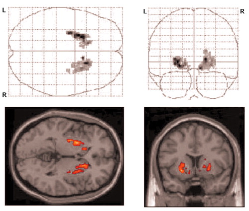
There was a significant difference in striatal [11C]raclopride binding potential between the balanced drink condition (mean=2.6, SD=0.3) and the tyrosine/phenylalanine depleted drink condition (mean=2.7, SD=0.3) (t=–5.10, df=6, p=0.002); dietary tyrosine/phenylalanine depletion increased [11C]raclopride binding potential in the striatum by a mean of 6% (SD=3%). The statistical parametric mapping analysis (Figure 1) confirmed the findings of the region of interest analysis. The simplified reference tissue model also showed a significant increase in binding potential (t=–3.6, df=6, p=0.01), as did analysis with fixed-volume regions of interest (t=–3.8, df=6, p=0.009). There was a significant increase in plasma prolactin concentration (an indirect measure of reduced dopamine transmission in the hypothalamus) from 4 hours before drink ingestion to 100 minutes after for the depleted drink condition (mean=177 [SD=56] versus 314 [SD=140], respectively) compared with the balanced drink condition (mean=184 [SD=53] and 145 [SD=39]) (F=5.86, df=2, 12, p<0.02). The change in ratio of plasma tyrosine and phenylalanine to large neutral amino acids was significantly greater for the depleted drink condition (tprescan: mean= 0.21 [SD=0.04]; tend: mean=0.0067 [SD=0.0038]) than for the balanced drink condition (tprescan: mean=0.23 [SD=0.03]; tend: mean=0.12 [SD=0.03]) (F=55.37, df=2, 12, p<0.001). The absolute concentration of tyrosine and phenylalanine was also lower relative to baseline in the depleted drink condition (–74%, SD=9) than in the balanced drink condition (266%, SD=147) (F=22.67, df=2, 12, p<0.001). Most importantly, the increase in binding potential following tyrosine and phenylalanine depletion correlated significantly with the percentage fall in the ratio of tyrosine and phenylalanine to large neutral amino acids (r=0.79, df=5, p=0.03), providing validation that the increase of [11C]raclopride binding was related to dopamine precursor availability.
Reference tissue time activity curve, region of interest volume, and radiopharmaceutical parameters (total injected, precursor injected, purity, and specific radioactivity) did not differ between conditions (data not shown).
Discussion
PET and microdialysis studies in primates have confirmed that changes in [11C]raclopride binding are associated with alterations in extracellular dopamine concentrations. Limited studies in nonhuman primates after administration of the dopamine-depleting agent alpha-methyl-para-tyrosine would indicate that an approximate 50% reduction in striatal extracellular dopamine concentration (measured with microdialysis) corresponds to a 25% increase in binding of the SPET radiotracer [123I]IBZM (6). This would indicate that the 6% increase in [11C]raclopride binding potential we report here may correspond to at least a 10%–20% reduction in dopamine concentrations. Indeed, because the balanced mixture also appeared to lower tyrosine availability to the brain to a moderate extent, greater increases of [11C]raclopride binding may have been seen in comparison with a control drink that did not alter ratios of tyrosine and phenylalanine to large neutral amino acids.
The reduction in presynaptic dopamine release produced by tyrosine and phenylalanine depletion offers the potential to lower the activity of dopamine pathways through a novel mechanism. Such an action could produce beneficial effects for a variety of neuropsychiatric syndromes in which clinical symptoms have been linked to increased dopamine release, such as acute schizophrenia and mania. Most conventional antipsychotic drugs are fairly selective antagonists of the D2 receptor subtype of dopamine receptor, whereas tyrosine and phenylalanine depletion would presumably lower neurotransmission at all postsynaptic dopamine receptor subtypes. In addition, the effect of diminished tyrosine availability to lower dopamine synthesis might be particularly apparent in those dopamine neurons that are firing most rapidly, perhaps resulting in the possibility of relatively selective inhibition of overactive pathways.
Received Sept. 20, 2002; revision received Feb. 4, 2003; accepted Feb. 6, 2003. From the Imperial College School of Medicine and the University Department of Psychiatry, Warneford Hospital, Oxford, U.K. Address reprint requests to Dr. Grasby, Imperial College School of Medicine, The Cyclotron Unit, Hammersmith Campus, DuCane Rd., W12 0NN, London, U.K.; [email protected] (e-mail). Supported by the Wellcome Trust (Dr. Montgomery) and the Medical Research Council (Drs. McTavish, Cowen, and Grasby).

Figure 1. Difference in [11C]Raclopride Binding in Seven Healthy Subjects After Ingestion of a Balanced Versus a Tyrosine/Phenylalanine-Depleted Amino Acid Drinka
aThe upper row shows axial and coronal statistical parametric mapping projections of the binding difference in the striatal region of interest. The lower row shows these regions projected onto representative magnetic resonance image brain slices.
1. Moja EA, Lucini V, Benedetti F, Lucca A: Decrease in plasma phenylalanine and tyrosine after phenylalanine-tyrosine free amino acid solutions in man. Life Sci 1996; 58:2389–2395Crossref, Medline, Google Scholar
2. McTavish SF, Cowen PJ, Sharp T: Effect of a tyrosine-free amino acid mixture on regional brain catecholamine synthesis and release. Psychopharmacology (Berl) 1999; 141:182–188Crossref, Medline, Google Scholar
3. Harmer CJ, McTavish SF, Clark L, Goodwin GM, Cowen PJ: Tyrosine depletion attenuates dopamine function in healthy volunteers. Psychopharmacology (Berl) 2001; 154:105–111Crossref, Medline, Google Scholar
4. McTavish SF, McPherson MH, Harmer CJ, Clark L, Sharp T, Goodwin GM, Cowen PJ: Antidopaminergic effects of dietary tyrosine depletion in healthy subjects and patients with manic illness. Br J Psychiatry 2001; 179:356–360Crossref, Medline, Google Scholar
5. Watabe H, Endres CJ, Breier A, Schmall B, Eckelman WC, Carson RE: Measurement of dopamine release with continuous infusion of [11C]raclopride: optimization and signal-to-noise considerations. J Nucl Med 2000; 41:522–530Medline, Google Scholar
6. Laruelle M, Iyer RN, al-Tikriti MS, Zea-Ponce Y, Malison R, Zoghbi SS, Baldwin RM, Kung HF, Charney DS, Hoffer PB, Innis RB, Bradberry CW: Microdialysis and SPECT measurements of amphetamine-induced dopamine release in nonhuman primates. Synapse 1997; 25:1–14Crossref, Medline, Google Scholar


