Progressive Loss of Cerebellar Volume in Childhood-Onset Schizophrenia
Abstract
OBJECTIVE: Childhood-onset schizophrenia is a severe and unremitting form of the disorder. Prospective brain magnetic resonance imaging (MRI) studies have found progressive loss of total cerebral volume during adolescence, primarily attributable to accelerated loss of cortical gray matter. Because there is evidence of cerebellar involvement in schizophrenia, the authors examined cerebellar volume and its relation to cortical gray matter development during adolescence in patients with childhood-onset schizophrenia and healthy comparison subjects. METHOD: Total cerebellar volume was algorithmically calculated for 108 anatomical brain MRI scans from 50 patients (20 of whom were female) and 101 scans from 50 age- and gender-matched healthy volunteers (20 of whom were female). The age range of the patients and comparison subjects was 8 to 24. Midsagittal vermal area and posterior-inferior vermal lobe volume were measured by hand. Prospective rescans were obtained at approximately 2-year intervals. Cross-sectional and longitudinal data were combined in mixed model regressions to compare developmental changes for the groups. RESULTS: In contrast to healthy volunteers, patients with schizophrenia showed a progressive loss of cerebellar volume during adolescence. Cerebellar and cerebral volume decreases were significantly correlated in childhood-onset schizophrenia. CONCLUSIONS: Childhood-onset schizophrenia is associated with significant progressive loss of cerebellar volume during adolescence, consistent with previously reported decreases in total cerebral and cortical gray matter. At least in these patients with severe early-onset schizophrenia, the loss appears secondary to a generalized process.
Childhood-onset schizophrenia, defined as onset of psychosis by age 12, is clinically and neurobiologically continuous with later-onset schizophrenia (1). Patients with this rare, severe illness have profound impairment in premorbid development (2) and resemble adult patients with poor-outcome schizophrenia. Previous National Institute of Mental Health (NIMH) brain imaging studies of childhood-onset schizophrenia reported progressive decreases in total volume in the cerebrum, hippocampus (3), and frontal and temporal cortical gray matter (4).
Cerebellar abnormalities have been reported, albeit inconsistently, in adult patients with schizophrenia. Some postmortem studies found smaller anterior vermal lobe with Purkinje cell dropout and thinning of granular and molecular layers (5) as well as reduction in linear density of vermal Purkinje cells (6), although others found no abnormalities (7, 8). Similarly, early computed tomography studies reported cerebellar atrophy (9, 10) and smaller cerebellar vermis (11–13), but quantitative magnetic resonance imaging (MRI) studies have produced inconsistent results (reviewed in references 14 and 15). Positive findings include reduced hemisphere and vermal volumes (16–18) and larger vermal white matter volume (19), but other MRI studies have shown no significant differences between patients with schizophrenia and comparison subjects (20–23). Studies of midsagittal vermal area in schizophrenia found larger area of lobules VI–VII (24), smaller anterior vermal lobe and smaller total vermis area (25, 26), or no abnormalities (27–29). Finally, one prospective MRI study reported significantly reduced right cerebellar volume in schizophrenia (30). Differences in subject selection, image acquisition, image analysis, and definition of boundaries make comparisons across studies difficult.
In a previous cross-sectional MRI study of 24 patients with childhood-onset schizophrenia, we found reduced vermal midsagittal area and posterior-inferior lobe volume (31). A subset of unmedicated adolescents with childhood-onset schizophrenia performing an auditory continuous performance task during positron emission tomography had greater than normal cerebellar glucose metabolism (32). Because prospective MRI studies are more sensitive to developmental change (33), the present study examined prospective anatomical brain scans during adolescence. Total cerebellar and posterior-inferior vermal volumes and the vermal midsagittal area were measured in patients with childhood-onset schizophrenia and healthy volunteers. We hypothesized that a decrement in cerebellar and posterior-inferior vermal volumes would be seen throughout adolescence. We were also interested in whether cerebellar changes paralleled previously reported cortical gray matter loss for this group.
Method
Childhood-Onset Schizophrenia
Children and adolescents were recruited nationally for an ongoing study of childhood-onset schizophrenia (34). Inclusion criteria were a DSM-III-R or DSM-IV diagnosis of schizophrenia with onset of psychosis before age 12, a premorbid full-scale IQ of at least 70, the absence of active medical or neurological disease, and poor response to or inability to tolerate treatment with at least two different neuroleptics. The diagnosis was established by using previous records and clinical and structured interviews of the children and parents as described in detail elsewhere (35).
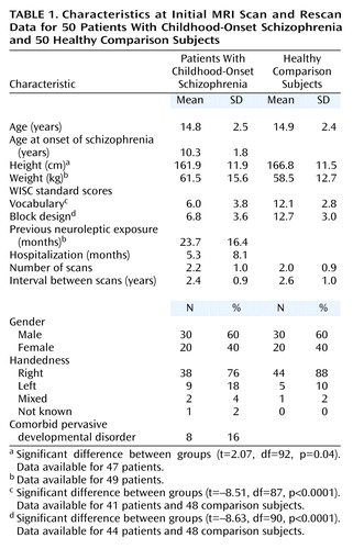
The study group of 50 patients with childhood-onset schizophrenia for whom at least one scan was available included 20 females. At baseline and at each follow-up visit, subjects were administered the WISC-R, the WISC-III, or the WAIS-R as appropriate. At follow-up, WISC raw scores for information and comprehension subscales were obtained to compare performance on these scales independent of subject age. A previously described method (2) was used to review premorbid neuropsychological, school, and medical records. Twenty-seven patients (nine of whom were female) had developmental language difficulties, and nine had comorbid pervasive developmental disorder, not otherwise specified, according to the Autism Screening Questionnaire (36).
Analyses of initial scans included 50 scans per diagnostic group. Patients returned for rescan approximately every 2 years. In the childhood-onset schizophrenia group, 19 patients had two, 12 had three, and five had four scans. In the comparison group, 19 subjects had two, 13 had three, and two had four scans. Developmental analyses were based on 108 childhood-onset schizophrenia scans and 101 comparison group scans. Using only subjects with more than one scan, we examined the relationship between cerebellar volume change and clinical or anatomical measures for 34 healthy volunteers and 36 patients with childhood-onset schizophrenia. All patients with childhood-onset schizophrenia were taking neuroleptics at follow-up, most frequently a combination of clozapine and at least one other antipsychotic medication (N=14), clozapine alone (N=12), or antipsychotic drugs other than clozapine (N=10).
Healthy Volunteers
Fifty healthy children and adolescents matched for age, sex, and handedness were recruited from the community (Table 1). Structured rating scales and interviews of child and parents were performed as described elsewhere (37).
Assent from the child and written consent from the parents were obtained for both patients and comparison subjects. The NIMH Institutional Review Board approved this study.
MRI Image Acquisition
All subjects were scanned on the same GE 1.5-T Signa scanner (GE Medical Systems, Milwaukee). T1-weighted images with contiguous 1.5-mm slices in the axial plane and 2.0-mm slices in the coronal plane were obtained by using three-dimensional spoiled gradient recalled echo in the steady state. Imaging parameters were echo time=5 msec, repetition time=24 msec, flip angle=45°, acquisition matrix=192×256, number of excitations=1, and field of view=24 cm. Head placement was standardized as previously described (3).
Image Analysis
Cerebrum and cerebellum
Total cerebral and cerebellar volumes were quantified by using a three-part fully automated image analysis process described in detail elsewhere (33, 38). Reliability of the algorithms equaled unity for test-retest with the same scan. Intraclass correlation coefficients (ICC) for 10 subjects rescanned after leaving and reentering the magnet exceeded 0.98 for both measures. Automated and hand-traced measures of cerebellar volume were highly reliable (N=17, ICC=0.94).
Vermis
Midsagittal vermal area and posterior-inferior vermal lobe volume, excluding the cerebellar tonsils, were hand-measured by one rater (A.C.V.) using the MNI Display program (Montreal Neurological Institute, McGill University, Montreal) on a Silicon Graphics workstation (Mountain View, Calif.). Beginning with the cerebral midsagittal slice, the best vermis midsagittal slice, which differed from the midcerebral 63% of the time (39), was selected in sagittal view and confirmed in coronal and axial views (Figure 1). Areas for anterior, superior-inferior, and posterior-inferior lobes were hand-traced on the vermis midsagittal slice and added to derive total vermis area. Boundaries for the anterior lobe are the primary fissure and fourth ventricle; boundaries for the superior-inferior lobe were the primary and prepyramidal fissures; boundaries for the posterior-inferior lobe (lobules VIII–X) were the prepyramidal fissure and fourth ventricle (31, 40).
Posterior-inferior volume was obtained by starting with the midsagittal outline that provided superior and inferior landmarks on each coronal slice. Lateral boundaries for each coronal slice were the CSF-gray matter interface (Figure 1), and filled coronal areas were checked slice by slice in axial and sagittal views. Measurement of volumes of anterior and superior-anterior vermis was not attempted because these regions do not have true lateral boundaries (40).
Interrater reliability between primary (A.C.V.) and secondary (A.K.) raters was obtained twice to check for rater drift with at least 10 brains per run; mean ICC=0.94 (SD=0.06) for posterior-inferior volume, and mean ICC=0. 93 (SD=0.06) for vermal area. Intrarater reliability was verified on three occasions: mean ICC=0.87 (SD=0.07) for posterior-inferior volume, and mean ICC=0.93 (SD=0.02) for vermal area.
Statistical Analysis
Fisher’s exact and Mann-Whitney test procedures were used to evaluate the comparability of patient and comparison groups.
Polynomial growth models were used to examine growth patterns of brain structures for initial (cross-sectional) scans. The initial cubic model was size=intercept + beta1* (age–mean age) + beta2* (age–mean age)2 + beta3* (age–mean age)3 + ε.
The model parameters (intercept and beta coefficients) were initially allowed to vary by sex and diagnostic group. The full cubic model was compared with simpler quadratic, linear, and constant models. Once the order of the model was established, testing was performed to determine whether an additive model could replace the interactions between sex and diagnostic group for the height and shape parameters of the curves. Hypothesis tests and model selection were initially based on F statistics. For F statistics with p values less than 0.10 that related to group or sex differences, permutation tests were performed to lessen the likelihood that a significant finding was due to a small number of outliers. To implement this procedure, 500 analyses using the preferred model were performed in which the sex and diagnostic group designation were randomly reassigned. The original F statistic based on the correct group and sex designation was compared with the resulting 500 F statistics, and an empirical p value was obtained.
The same polynomial growth models were used for the analysis of longitudinal data including all of each individual’s scans (41, 42). To account for within-person correlation, intercepts were treated as normally distributed random effects that varied by individual, while beta coefficients for age, age-squared, and age-cubed terms were modeled as fixed effects. Although the statistical information provided by individuals with only one scan may be less than that obtained from those with multiple scans, single scans do provide additional information about between-person variation and overall curve shape.
Spearman correlation was used to examine the relationship between slopes for cerebellar and total cerebral volume loss. Slopes were calculated only for subjects with more than one scan; for those with more than two scans, first and last scans were selected. Stepwise regression and analysis of variance were used to examine clinical and treatment variables in relation to cerebellar volume decrease in childhood-onset schizophrenia.
Results
Initial Scans
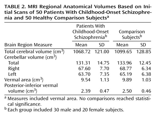
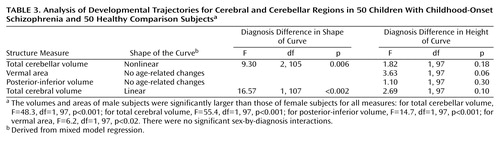
As expected, all volumes were greater for males than females (p<0.02) (Table 2 and Table 3). In analyses limited to each subject’s initial scan, no significant diagnostic differences (Table 2) or sex-by-diagnosis interactions were seen for childhood-onset schizophrenia and healthy comparison subjects for total cerebral volume, cerebellar measures, or vermal measures.
Developmental Trajectories
Total cerebellar volume
In contrast, as seen in Table 3 and Figure 2, developmental trajectories differed significantly for the childhood-onset schizophrenia group (F=9.30, df=2, 105, p=0.006), with total cerebellar volume decreasing with increasing age for patients with childhood-onset schizophrenia but not for comparison subjects. For comparison purposes, and in agreement with previous reports (3), developmental trajectories for total cerebral volume also differed significantly for the childhood-onset schizophrenia group (F=16.57, df=1, 107, p<0.002). As seen in Figure 2, total cerebral volume decreased with age for patients with childhood-onset schizophrenia but not for healthy comparison subjects. By visual examination, losses of cerebellar and cerebral volumes appeared to start at approximately the same time.
Vermal area and posterior-inferior vermal volume
No age-related changes were found for either comparison subjects or patients with childhood-onset schizophrenia.
Cerebellar volume change and anatomical and clinical measures
Using only childhood-onset schizophrenia subjects with more than one scan, we found that rates (cm3/year) of decrease of total cerebellar and cerebral volumes were significantly correlated (r=0.41, N=36, p=0.01). Within the patient group, no significant relationships were observed between cerebellar volume decrease and scores on WISC vocabulary or block design subscales; age at onset; number of months of hospitalization; history of prepsychotic language disorder; pervasive developmental disorder diagnosis; scores on the Scale for the Assessment of Negative Symptoms, Scale for the Assessment of Positive Symptoms, or Brief Psychiatric Rating Scale; medication group (clozapine alone, clozapine with at least one other antipsychotic drug, other antipsychotic); or slopes for change in full-scale IQ or in information and comprehension subscale raw scores.
Discussion
In this prospective study of cerebellar morphology in childhood-onset schizophrenia, a progressive loss of cerebellar volume in childhood-onset schizophrenia was found during adolescence, paralleling the loss in total cerebral volume previously reported for this group (3). The cerebellum is one of the first brain structures to begin to differentiate and one of the last to achieve maturity (43), reaching peak volume several years later than total cerebral volume (Giedd et al., unpublished data). However, in patients with childhood-onset schizophrenia, cerebellar loss seems to start at about the same age (within 1 year, by visual examination of confidence intervals) as loss of cerebral volume (Figure 2). The fact that cerebellar tissue loss appears simultaneously with that of the total cerebrum suggests a generalized abnormal process in childhood-onset schizophrenia superimposed on normal development.
When only initial scans were examined, no difference in cerebellar volume was seen between the 50 patients with childhood-onset schizophrenia and the 50 normal comparison subjects. As with other measures in patients with childhood-onset schizophrenia and healthy subjects, only the availability of prospective longitudinal data revealed subtle differences in developmental trajectories (3, 33) that were not detectable in cross-sectional studies with the same subjects (37, 44).
These findings are consistent with reported abnormal cerebellar function in childhood and adult schizophrenia (32, 45–47). There is accumulating evidence for a cognitive role for the cerebellum (see review in reference 48), including executive function and working memory, which are impaired in schizophrenia (49–53). Positron emission tomography studies have revealed abnormalities in cerebellar blood flow while patients carry out cognitive tasks (46, 47). A model of schizophrenia as secondary to disrupted development in a cortico-cerebellar-thalamic-cortical circuit (46) has been termed “cognitive dysmetria” (54, 55), referring to incoordination in the processing, prioritization, retrieval, and expression of information. Unlike a previous study (56), however, we found no significant relationship between any symptom pattern or change in any clinical or cognitive measure and our measures of cerebellar volume loss. We did not have available measures of motor function, which might have revealed a relationship to cerebellar volume loss.
We did not replicate previous findings of decreased midsagittal vermal area and posterior-inferior vermal lobe volume (31), possibly due to the vagaries of hand measurements of small structures. In spite of good intrarater and interrater reliabilities, the anatomy of the vermis is difficult to delineate (40), rater measures are subject to drift, and techniques for hand-measuring are fraught with methodological issues such as location of vermal midslice (39, 57, 58).
Limitations of the study include substantially lower IQ for the patient group. Moreover, since all of our patients were medicated, we cannot rule out the possibility that our findings reflect the effects of neuroleptic medications. Our focus on treatment-resistant subjects also limits the generalizability of these findings.
In summary, the excess loss of brain tissue during adolescence seen in this and other studies may be a trait marker for schizophrenia. Since there is evidence of reduction of cerebellar volume in relatives of adult patients with schizophrenia compared with control subjects (59), we are currently conducting a prospective brain MRI study of the siblings of our patients.
 |
 |
 |
Received Dec. 11, 2001; revision received June 26, 2002; accepted July 8, 2002. From the Child Psychiatry Branch, National Institute of Mental Health. Address reprint requests to Dr. Rapoport, Child Psychiatry Branch, National Institute of Mental Health, Bldg. 10, Rm. 3N 202, Bethesda, MD 20892-1600; [email protected] (e-mail). The author thank Prof. François Ansermet and the National Swiss Research Fund for their support to Dr. Keller, and Jonathan Blumenthal, Liv S. Clasen, and Peter Gochman for technical assistance.

Figure 1. Posterior-Inferior Vermis Methodological Outlines Used for MRI Scans of 50 Patients With Childhood-Onset Schizophrenia and 50 Healthy Comparison Subjectsa
aThe best vermis midsagittal slice was selected in sagittal view and confirmed in coronal and axial views with the aid of the crosshair cursor. Posterior-inferior volume was obtained by starting with the midsagittal outline that provided superior and inferior landmarks on each coronal slice. Lateral boundaries for each coronal slice were the interface between CSF and gray matter, and filled coronal areas were checked slice by slice in axial and sagittal views.

Figure 2. Total Cerebellar and Total Cerebral Volumes in Relation to Age for 108 MRI Scans of 50 Patients With Childhood-Onset Schizophrenia and 101 Scans of 50 Healthy Volunteers
1. Jacobsen LK, Rapoport JL: Research update: childhood-onset schizophrenia: implications of clinical and neurobiological research. J Child Psychol Psychiatry 1998; 39:101-113Crossref, Medline, Google Scholar
2. Nicolson R, Lenane M, Singaracharlu S, Malaspina D, Giedd JN, Hamburger SD, Gochman P, Bedwell J, Thaker GK, Fernandez T, Wudarsky M, Hommer DW, Rapoport JL: Premorbid speech and language impairments in childhood-onset schizophrenia: association with risk factors. Am J Psychiatry 2000; 157:794-800Link, Google Scholar
3. Giedd JN, Jeffries NO, Blumenthal J, Castellanos FX, Vaituzis AC, Fernandez T, Hamburger SD, Liu H, Nelson J, Bedwell J, Tran L, Lenane M, Nicolson R, Rapoport JL: Childhood-onset schizophrenia: progressive brain changes during adolescence. Biol Psychiatry 1999; 46:892-898Crossref, Medline, Google Scholar
4. Rapoport JL, Giedd JN, Blumenthal J, Hamburger S, Jeffries N, Fernandez T, Nicolson R, Bedwell J, Lenane M, Zijdenbos A, Paus T, Evans A: Progressive cortical change during adolescence in childhood-onset schizophrenia: a longitudinal magnetic resonance imaging study. Arch Gen Psychiatry 1999; 56:649-654Crossref, Medline, Google Scholar
5. Weinberger DR, Kleinman JE, Luchins DJ, Bigelow LB, Wyatt RJ: Cerebellar pathology in schizophrenia: a controlled postmortem study. Am J Psychiatry 1980; 137:359-361Link, Google Scholar
6. Reyes MG, Gordon A: Cerebellar vermis in schizophrenia. Lancet 1981; 2:700-701Crossref, Medline, Google Scholar
7. Supprian T, Ulmar G, Bauer M, Schuler M, Puschel K, Retz-Junginger P, Schmitt HP, Heinsen H: Cerebellar vermis area in schizophrenic patients—a post-mortem study. Schizophr Res 2000; 42:19-28Crossref, Medline, Google Scholar
8. Lohr JB, Jeste DV: Cerebellar pathology in schizophrenia? a neuronometric study. Biol Psychiatry 1986; 21:865-875Crossref, Medline, Google Scholar
9. Dewan MJ, Pandurangi AK, Lee SH, Ramachandran T, Levy BF, Boucher M, Yozawitz A, Major L: Cerebellar morphology in chronic schizophrenic patients: a controlled computed tomography study. Psychiatry Res 1983; 10:97-103Crossref, Medline, Google Scholar
10. Boronow J, Pickar D, Ninan PT, Roy A, Hommer D, Linnoila M, Paul SM: Atrophy limited to the third ventricle in chronic schizophrenic patients: report of a controlled series. Arch Gen Psychiatry 1985; 42:266-271Crossref, Medline, Google Scholar
11. Lippmann S, Manshadi M, Baldwin H, Drasin G, Rice J, Alrajeh S: Cerebellar vermis dimensions on computerized tomographic scans of schizophrenic and bipolar patients. Am J Psychiatry 1982; 139:667-668Link, Google Scholar
12. Heath RG, Franklin DE, Walker CF, Keating JW Jr: Cerebellar vermal atrophy in psychiatric patients. Biol Psychiatry 1982; 17:569-583Medline, Google Scholar
13. Sandyk R, Kay SR, Merriam AE: Atrophy of the cerebellar vermis: relevance to the symptoms of schizophrenia. Int J Neurosci 1991; 57:205-212Crossref, Medline, Google Scholar
14. McCarley RW, Wible CG, Frumin M, Hirayasu Y, Levitt JJ, Fischer IA, Shenton ME: MRI anatomy of schizophrenia. Biol Psychiatry 1999; 45:1099-1119Crossref, Medline, Google Scholar
15. Shenton ME, Dickey CC, Frumin M, McCarley RW: A review of MRI findings in schizophrenia. Schizophr Res 2001; 49:1-52Crossref, Medline, Google Scholar
16. Volz H, Gaser C, Sauer H: Supporting evidence for the model of cognitive dysmetria in schizophrenia—a structural magnetic resonance imaging study using deformation-based morphometry. Schizophr Res 2000; 46:45-56Crossref, Medline, Google Scholar
17. Ichimiya T, Okubo Y, Suhara T, Sudo Y: Reduced volume of the cerebellar vermis in neuroleptic-naive schizophrenia. Biol Psychiatry 2001; 49:20-27Crossref, Medline, Google Scholar
18. Loeber RT, Cintron CMB, Yurgelun-Todd DA: Morphometry of individual cerebellar lobules in schizophrenia. Am J Psychiatry 2001; 158:952-954Link, Google Scholar
19. Levitt JJ, McCarley RW, Nestor PG, Petrescu C, Donnino R, Hirayasu Y, Kikinis R, Jolesz FA, Shenton ME: Quantitative volumetric MRI study of the cerebellum and vermis in schizophrenia: clinical and cognitive correlates. Am J Psychiatry 1999; 156:1105-1107Abstract, Google Scholar
20. Andreasen NC, Flashman L, Flaum M, Arndt S, Swayze V II, O’Leary DS, Ehrhardt JC, Yuh WTC: Regional brain abnormalities in schizophrenia measured with magnetic resonance imaging. JAMA 1994; 272:1763-1769Crossref, Medline, Google Scholar
21. Flaum M, Swayze VW II, O’Leary DS, Yuh WTC, Ehrhardt JC, Arndt SV, Andreasen NC: Effects of diagnosis, laterality, and gender on brain morphology in schizophrenia. Am J Psychiatry 1995; 152:704-714Link, Google Scholar
22. Keshavan MS, Haas GL, Kahn CE, Aguilar E, Dick EL, Schooler NR, Sweeney JA, Pettegrew JW: Superior temporal gyrus and the course of early schizophrenia: progressive, static, or reversible? J Psychiatr Res 1998; 32:161-167Crossref, Medline, Google Scholar
23. Staal WG, Hulshoff Pol HE, Schnack HG, Hoogendoorn MLC, Jellema K, Kahn RS: Structural brain abnormalities in patients with schizophrenia and their healthy siblings. Am J Psychiatry 2000; 157:416-421Link, Google Scholar
24. Nasrallah HA, Schwarzkopf SB, Olson SC, Coffman JA: Perinatal brain injury and cerebellar vermal lobules I-X in schizophrenia. Biol Psychiatry 1991; 29:567-574Crossref, Medline, Google Scholar
25. Nopoulos PC, Ceilley JW, Gailis EA, Andreasen NC: An MRI study of cerebellar vermis morphology in patients with schizophrenia: evidence in support of the cognitive dysmetria concept. Biol Psychiatry 1999; 46:703-711Crossref, Medline, Google Scholar
26. Rossi A, Stratta P, Mancini F, Decataldo S, Casacchia M: Cerebellar vermal size in schizophrenia—a male effect. Biol Psychiatry 1993; 33:354-357Crossref, Medline, Google Scholar
27. Mathew RJ, Partain CL: Midsagittal sections of the cerebellar vermis and fourth ventricle obtained with magnetic resonance imaging of schizophrenic patients. Am J Psychiatry 1985; 142:970-971Link, Google Scholar
28. Aylward EH, Reiss A, Barta PE, Tien A, Han W, Lee J, Pearlson GD: Magnetic resonance imaging measurement of posterior fossa structures in schizophrenia. Am J Psychiatry 1994; 151:1448-1452Link, Google Scholar
29. Uematsu M, Kaiya H: Midsagittal cortical pathomorphology of schizophrenia: a magnetic resonance imaging study. Psychiatry Res 1989; 30:11-20Crossref, Medline, Google Scholar
30. Delisi LE, Sakuma M, Tew W, Kushner M, Hoff AL, Grimson R: Schizophrenia as a chronic active brain process: a study of progressive brain structural change subsequent to the onset of schizophrenia. Psychiatry Res 1997; 74:129-140Crossref, Medline, Google Scholar
31. Jacobsen LK, Giedd JN, Berquin PC, Krain AL, Hamburger SD, Kumra S, Rapoport JL: Quantitative morphology of the cerebellum and fourth ventricle in childhood-onset schizophrenia. Am J Psychiatry 1997; 154:1663-1669Link, Google Scholar
32. Jacobsen LK, Hamburger SD, Van Horn JD, Vaituzis AC, McKenna K, Frazier JA, Gordon CT, Lenane MC, Rapoport JL, Zametkin AJ: Cerebral glucose metabolism in childhood onset schizophrenia. Psychiatry Res 1997; 75:131-144Crossref, Medline, Google Scholar
33. Giedd JN, Blumenthal J, Jeffries NO, Castellanos FX, Liu H, Zijdenbos A, Paus T, Evans AC, Rapoport JL: Brain development during childhood and adolescence: a longitudinal MRI study. Nat Neurosci 1999; 2:861-863Crossref, Medline, Google Scholar
34. Nicolson R, Rapoport JL: Childhood-onset schizophrenia: rare but worth studying. Biol Psychiatry 1999; 46:1418-1428Crossref, Medline, Google Scholar
35. Kumra S, Jacobsen LK, Lenane M, Karp BI, Frazier JA, Smith AK, Bedwell J, Lee P, Malanga CJ, Hamburger S, Rapoport JL: Childhood-onset schizophrenia: an open-label study of olanzapine in adolescents. J Am Acad Child Adolesc Psychiatry 1998; 37:377-385Crossref, Medline, Google Scholar
36. Berument SK, Rutter M, Lord C, Pickles A, Bailey A: Autism Screening Questionnaire: diagnostic validity. Br J Psychiatry 1999; 175:444-451Crossref, Medline, Google Scholar
37. Giedd JN, Snell JW, Lange N, Rajapakse JC, Casey BJ, Kozuch PL, Vaituzis AC, Vauss YC, Hamburger SD, Kaysen D, Rapoport JL: Quantitative magnetic resonance imaging of human brain development: ages 4-18. Cereb Cortex 1996; 6:551-560Crossref, Medline, Google Scholar
38. Collins L, Holmes CJ, Peters TM, Evans AC: Automatic 3D model-based neuroanatomical segmentation. Hum Brain Mapp 1995; 3:190-208Crossref, Google Scholar
39. Courchesne E, Press GA, Murakami J, Berthoty D, Grafe M, Wiley CA, Hesselink JR: The cerebellum in sagittal plane—anatomic-MR correlation, 1: the vermis. AJR Am J Roentgenol 1989; 153:829-835Crossref, Medline, Google Scholar
40. Schmahmann JD, Doyon J, McDonald D, Holmes C, Lavoie K, Hurwitz AS, Kabani N, Toga A, Evans A, Petrides M: Three-dimensional MRI atlas of the human cerebellum in proportional stereotaxic space. Neuroimage 1999; 10:233-260Crossref, Medline, Google Scholar
41. Diggle PJ, Liang KY, Zeger SL: Analysis of Longitudinal Data. Oxford, UK, Clarendon Press (Oxford University Press), 1994Google Scholar
42. Verbeke G, Molenberghs G: Linear Mixed Models in Practice: An SAS-Oriented Approach. New York, Springer Verlag, 1997Google Scholar
43. Wang VY, Zoghbi HY: Genetic regulation of cerebellar development. Nat Rev Neurosci 2001; 2:484-491Crossref, Medline, Google Scholar
44. Frazier JA, Giedd JN, Hamburger SD, Albus KE, Kaysen D, Vaituzis AC, Rajapakse JC, Lenane MC, McKenna K, Jacobsen LK, Gordon CT, Breier A, Rapoport JL: Brain anatomic magnetic resonance imaging in childhood-onset schizophrenia. Arch Gen Psychiatry 1996; 53:617-624Crossref, Medline, Google Scholar
45. Volkow ND, Levy A, Brodie JD, Wolf AP, Cancro R, Van Gelder P, Henn F: Low cerebellar metabolism in medicated patients with chronic schizophrenia. Am J Psychiatry 1992; 149:686-688Link, Google Scholar
46. Andreasen NC, O’Leary DS, Cizadlo T, Arndt S, Rezai K, Boles Ponto LL, Watkins GL, Hichwa RD: Schizophrenia and cognitive dysmetria: a positron-emission tomography study of dysfunctional prefrontal-thalamic-cerebellar circuitry. Proc Natl Acad Sci USA 1996; 93:9985-9990Crossref, Medline, Google Scholar
47. Andreasen NC, O’Leary DS, Flaum M, Nopoulos P, Watkins GL, Boles Ponto LL, Hichwa RD: Hypofrontality in schizophrenia: distributed dysfunctional circuits in neuroleptic-naive patients. Lancet 1997; 349:1730-1734Crossref, Medline, Google Scholar
48. Middleton FA, Strick PL: Basal ganglia and cerebellar loops: motor and cognitive circuits. Brain Res Brain Res Rev 2000; 31:236-250Crossref, Medline, Google Scholar
49. Goldberg TE, Gold JM, Greenberg R, Griffin S, Schulz SC, Pickar D, Kleinman JE, Weinberger DR: Contrasts between patients with affective disorders and patients with schizophrenia on a neuropsychological test battery. Am J Psychiatry 1993; 150:1355-1362Link, Google Scholar
50. Appollonio IM, Grafman J, Schwartz V, Massaquoi S, Hallett M: Memory in patients with cerebellar degeneration. Neurology 1993; 43:1536-1544Crossref, Medline, Google Scholar
51. Goldman-Rakic PS: Working memory dysfunction in schizophrenia. J Neuropsychiatry Clin Neurosci 1994; 6:348-357Crossref, Medline, Google Scholar
52. Bower JM: Control of sensory data acquisition. Int Rev Neurobiol 1997; 41:489-513Crossref, Medline, Google Scholar
53. Schmahmann JD, Sherman JC: The cerebellar cognitive affective syndrome. Brain 1998; 121:561-579Crossref, Medline, Google Scholar
54. Andreasen NC, Paradiso S, O’Leary DS: “Cognitive dysmetria” as an integrative theory of schizophrenia: a dysfunction in cortical-subcortical-cerebellar circuitry? Schizophr Bull 1998; 24:203-218Crossref, Medline, Google Scholar
55. Wiser AK, Andreasen NC, O’Leary DS, Watkins GL, Boles Ponto LL, Hichwa RD: Dysfunctional cortico-cerebellar circuits cause “cognitive dysmetria” in schizophrenia. Neuroreport 1998; 9:1895-1899Crossref, Medline, Google Scholar
56. Wassink TH, Andreasen NC, Nopoulos P, Flaum M: Cerebellar morphology as a predictor of symptom and psychosocial outcome in schizophrenia. Biol Psychiatry 1999; 45:41-48Crossref, Medline, Google Scholar
57. Coffman JA, Schwarzkopf SB, Olson SC, Nasrallah HA: Midsagittal cerebral anatomy by magnetic resonance imaging: the importance of slice position and thickness. Schizophr Res 1989; 2:287-294Crossref, Medline, Google Scholar
58. Courchesne E, Yeung-Courchesne R, Egaas B: Methodology in neuroanatomic measurement. Neurology 1994; 44:203-208Crossref, Medline, Google Scholar
59. Seidman LJ, Faraone SV, Goldstein JM, Goodman JM, Kremen WS, Toomey R, Tourville J, Kennedy D, Makris N, Caviness VS, Tsuang MT: Thalamic and amygdala-hippocampal volume reductions in first-degree relatives of patients with schizophrenia: an MRI-based morphometric analysis. Biol Psychiatry 1999; 46:941-954Crossref, Medline, Google Scholar