Brain Development, II: Establishing Synaptic Connections
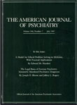
During normal CNS development, neurons in various brain regions express chemical markers that attract ingrowing neuronal terminals to form synapses with their correct targets. A single group of neurons within a neuronal structure may offer distinctive chemical cues that are organized into meaningful anatomic patterns to attract afferents from different brain regions. The cerebellum, for example, receives axons from spinal cord neurons whose terminals are distributed into distinct columns in the cerebellar cortex (bright pink areas in left figure). The terminals of spinocerebellar axons are labeled by injecting a dye, horseradish peroxidase, into the spinal cord, where it is taken up by neurons and transported to their terminal ends. The distinctive distribution of terminals in the cerebellum correlates with biochemical differences among cerebellar Purkinje cells. Thus, it is thought that the Purkinje cells express the chemical markers that organize the spinocerebellar terminals into distinct fields.
This model is being tested by studying a genetic strain of mice, staggerer, that fails to develop mature Purkinje cells. Staggerer (gene symbol, sg) is an autosomal recessive mutation that causes a block in the development of cerebellar Purkinje cells. The number of Purkinje cells is reduced by 75%, and the surviving Purkinje cells fail to differentiate completely. As a consequence of this Purkinje cell loss, the vast majority of granule cells (primary afferents to Purkinje cells) in the cerebellar cortex also die. If Purkinje cells are the organizers for cerebellar afferent projections, then in their absence the organization of spinocerebellar afferents should be disrupted. In older staggerer mice, the terminals of spinocerebellar axons are indeed reduced in density and lack any apparent organization. However, in juvenile staggerer mice (right figure), the afferent terminals still can be seen to form a pattern that is approximately normal, even in a cerebellum diminished by granule and Purkinje cell loss. The results suggest that even immature Purkinje cells may express the chemical markers that allow afferent axons to sort out into distinct columns. Modern molecular techniques have identified a growing number of transcription factors (DNA binding proteins such as Engrailed and Pax), nerve cell adhesion molecules (e.g., N-CAM), and signaling molecules (e.g., the semiphorins and collapsins) with distinct expression patterns in the developing nervous system. These molecules and the cells that express them are under intense study so that we can identify the molecular mechanisms that organize how the nervous system becomes wired together during development.
Address reprint requests to Dr. Tamminga, Maryland Psychiatric Research Center, P.O. Box 21247, Baltimore, MD 21228. Photographs courtesy of Dr. Vogel.

FIGURE 1. The horseradish-peroxidase-labeled terminals of spinocerebellar mossy fiber afferents (bright pink areas) segregate into discrete columns in the cerebellar cortex of wild type mice (left photomicrograph) and juvenile staggerer mutant mice (right photomicrograph), despite the defects in staggerer Purkinje cells and the loss of granule cells in this mutant. Scale bar, 200 µm.